

ADAPT-3D – The Future of Rapid Whole-Mount Tissue Clearing & Labeling
Master the 3rd Dimension: Accelerated Deep-Tissue Labeling with ADAPT-3D

Overcome the “Time Bottleneck” – Achieve high-fidelity 3D spatial insights in days, not weeks.
The spatial biology revolution is here, but your results are only as good as your sample prep. ADAPT-3D is a standardized, high-throughput protocol designed to solve the primary challenge of large-volume imaging: deep-tissue probe penetration.

The Challenge: Why 2D Histology is Leaving You Behind
Traditional sectioning destroys the spatial context of the cellular environment. However, current 3D methods like CLARITY or iDISCO often force a compromise: either you wait weeks for antibody diffusion, or you settle for “hollow” staining in the center of your sample.
The ADAPT-3D Difference:
We’ve optimized the chemical kinetics of delipidation and labeling. By managing the tissue’s physical state, ADAPT-3D drives antibodies deeper and faster, ensuring uniform signal intensity from the surface to the core.
Why ADAPT 3D Prevents Tissue Shrinkage
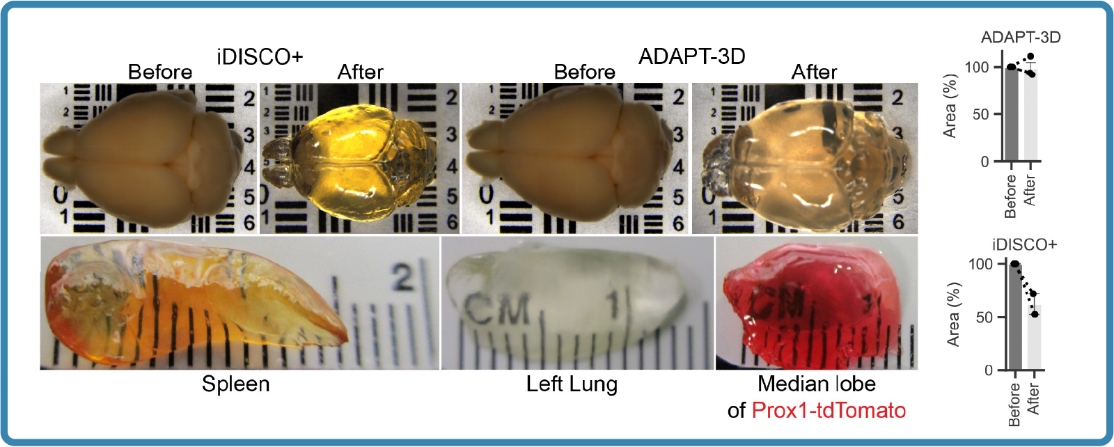
Traditional clearing methods (like BABB, DISCO, or even high-concentration sugar protocols) often cause significant tissue shrinkage—sometimes up to 30–50% of the original volume. This happens because high-molarity solvents or dehydrating agents pull water out of the cells too aggressively.
 Note: DCM-FREE FORMULA: This product is 100% free of Dichloromethane (Methylene Chloride). Formulated for user safety, FDA, and environmental compliance.
Engineered for Precision. Optimized for Speed.
Rapid Whole-Mount Labeling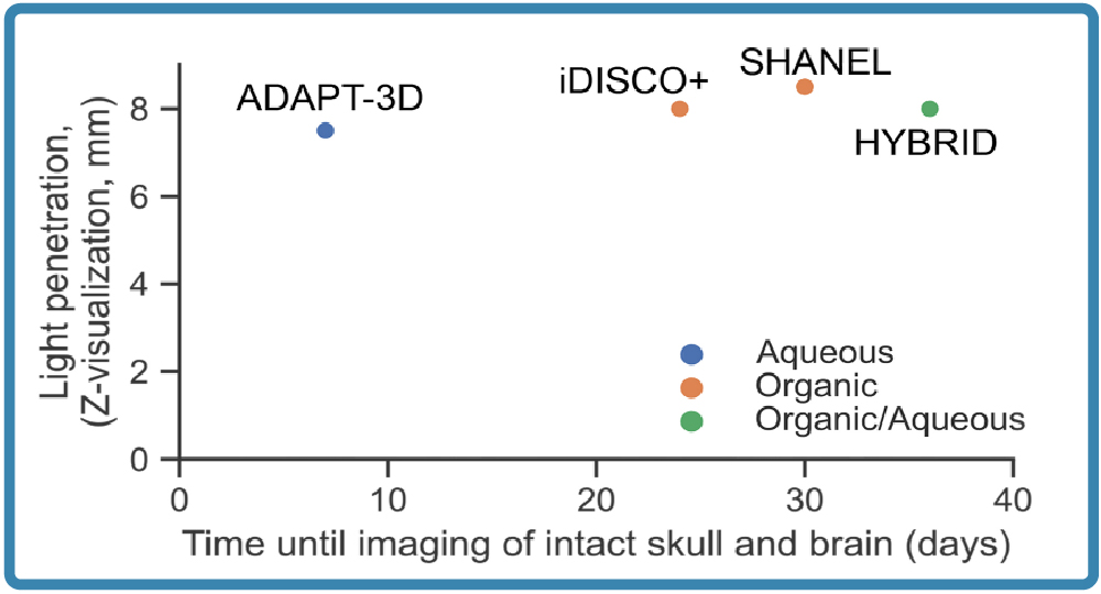
Why wait 14 days for a whole mouse brain? ADAPT-3D reduces incubation times to about 6 days, that’s up to 70% without compromising the structural integrity of the tissue.
Unmatched Signal-to-Noise
Specifically formulated to preserve endogenous fluorescence (GFP, YFP, tdTomato) while minimizing background autofluorescence.
Perfect Refractive Index (RI) Matching
Our protocol includes a custom RIMS (Refractive Index Matching Solution) workflow, achieving a perfect RI = 1.52, ideal for seamless Light Sheet and Confocal imaging.
“One-Stop” Compatibility
Whether you are using Xenium, PhenoCycler, or Light Sheet Fluorescence Microscopy (LSFM), ADAPT-3D integrates into your existing spatial omics workflow.
From Bench to Breakthrough: Applications
“ADAPT-3D represents the missing link in high-throughput 3D phenotyping. It allows us to see the ‘unseen’ within intact biological systems.” – Dr. Daniel D. Lee, Washington University
- Neuroscience: Map whole-brain neural circuits and synaptic densities in 3D.
- Oncology: Visualize the Tumor Microenvironment (TME) and immune cell infiltration in intact human biopsies.
- Immunology: Track lymphatic drainage and leukocyte trafficking through whole-mount organs.
- Drug Discovery: Perform 3D pharmacokinetic studies to see exactly where your drug candidates distribute in a tissue.
Individual Bulk Options Now Available
All ADAPT-3D components are now available for individual bulk purchase. This modular approach allows you to scale efficiently by ordering only the specific elements your project requires.
Technical Specifications & Workflow

