

Anti-Mouse PD-1 (CD279) [Clone 384-35] — Recombinant in vivo Functional Grade

Anti-Mouse PD-1 (CD279) [Clone 384-35] — Recombinant in vivo Functional Grade
Product No.: P710
- -
- -
Product No.P710 Clone 384-35 Target PD-1 Product Type Recombinant Monoclonal Antibody Alternate Names Programmed Death-1, CD279, PD1, PDCD1 Isotype Mouse IgG1 Applications B , FA , in vivo |
Data
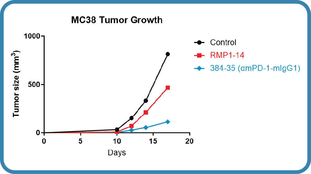 Administration of mAb 384-35 shows tumor volume more than 2.5 fold smaller compared to RMP1-14
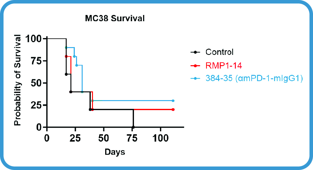 Administration of mAb 384-35 significantly increases survival past 100 days compared to RMP1-14
 - -
- -
Antibody DetailsProduct DetailsReactive Species Mouse Host Species Mouse FC Effector Activity Active Recommended Isotype Controls Immunogen Mouse PD-1 Product Concentration ≥ 5.0 mg/ml Endotoxin Level <1.0 EU/µg Purity ≥95% monomer by analytical SEC ⋅ >95% by SDS Page Formulation This recombinant monoclonal antibody is aseptically packaged and formulated in 0.01 M phosphate buffered saline (150 mM NaCl) PBS pH 7.2 - 7.4 with no carrier protein, potassium, calcium or preservatives added. Due to inherent biochemical properties of antibodies, certain products may be prone to precipitation over time. Precipitation may be removed by aseptic centrifugation and/or filtration. State of Matter Liquid Product Preparation Functional grade preclinical antibodies are manufactured in an animal free facility using in vitro cell culture techniques and are purified by a multi-step process including the use of protein A or G to assure extremely low levels of endotoxins, leachable protein A or aggregates. Pathogen Testing To protect mouse colonies from infection by pathogens and to assure that experimental preclinical data is not affected by such pathogens, all of Leinco’s recombinant antibodies are tested and guaranteed to be negative for all pathogens in the IDEXX IMPACT I Mouse Profile. Storage and Handling Functional grade preclinical antibodies may be stored sterile as received at 2-8°C for up to one month. For longer term storage, aseptically aliquot in working volumes without diluting and store at ≤ -70°C. Avoid Repeated Freeze Thaw Cycles. Regulatory Status Research Use Only (RUO). Non-Therapeutic. Country of Origin USA Shipping 2-8°C Wet Ice Additional Applications Reported In Literature ? B, FA, in vivo, Each investigator should determine their own optimal working dilution for specific applications. See directions on lot specific datasheets, as information may periodically change. DescriptionDescriptionSpecificity Anti-PD1 antibody (clone 384-35) recognizes an epitope on mouse PD-1. Background This anti-PD1 antibody was generated by immunizing PD-1-/- mice with mouse PD-1. The resulting antibody is fully murinized and contains no sequences from other species.
PD-1 is a 50-55 kD member of the B7 Ig superfamily. PD-1 is also a member of the extended CD28/CTLA-4 family of T cell regulators and is suspected to play a role in lymphocyte clonal selection and peripheral tolerance. The ligands of PD-1 are PD-L1 and PD-L2, and are also members of the B7 Ig superfamily. PD-1 and its ligands negatively regulate immune responses. PD-L1, or B7-Homolog 1, is a 40 kD type I transmembrane protein that has been reported to costimulate T cell growth and cytokine production. The interaction of PD-1 with its ligand PD-L1 is critical in the inhibition of T cell responses that include T cell proliferation and cytokine production. PD-L1 has increased expression in several cancers. Inhibition of the interaction between PD-1 and PD-L1 can serve as an immune checkpoint blockade by improving T-cell responses In vitro and mediating preclinical antitumor activity. Within the field of checkpoint inhibition, combination therapy using anti-PD1 in conjunction with anti-CTLA4 has significant therapeutic potential for tumor treatments. PD-L2 is a 25 kD type I transmembrane ligand of PD-1. Via PD-1, PD-L2 can serve as a co-inhibitor of T cell functions. Regulation of T cell responses, including enhanced T cell proliferation and cytokine production, can result from mAbs that block the PD-L2 and PD-1 interaction. Antigen Distribution PD-1 is expressed on a subset of CD4-CD8- thymocytes, and on activated T and B cells. Ligand/Receptor PD-L1 (B7-H1), PD-L2 PubMed NCBI Gene Bank ID UniProt.org Q02242 Research Area Apoptosis . Cancer . Cell Biology . Cell Death . Immunology . Inhibitory Molecules . Tumor Suppressors References & Citations1. Schreiber, RD. et al. (2017) Cancer Immunol Res. 5(2):106-117. 2. Honjo, T. et al. (1992) EMBO J. 11:3887. |
Formats Available
- -
- -
Prod No. | Description |
|---|---|
P710 |
