

Immune Checkpoint Signaling Pathways
Immune checkpoints serve as crucial molecules that modulate the behavior of immune cells. Often, immune checkpoints are mediated via T cell surface proteins that must bind to partner receptor proteins on other cells (e.g., antigen-presenting cells or APCs).
To perform its normal function, the immune system must distinguish between normal and abnormal cells. In the best case, healthy cells remain intact, while infected or cancerous cells are attacked and destroyed. For this process, leukocytes use immune checkpoints.
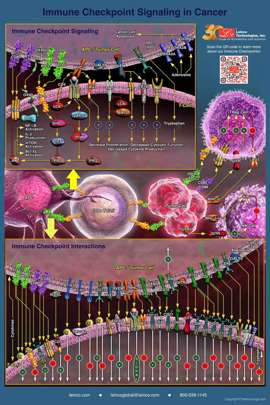
When the checkpoint and partner protein bind to each other, they send a signal that causes the immune system to perform a specific action such as proliferation or destruction of target cells. Activation of immune checkpoint signaling pathways can lead to either suppression or stimulation of the immune system, depending on the signaling pathway.
Co-stimulatory receptors include CD28 and ICOS (inducible T cell co-stimulator), 4-1BB, OX40, CD27, CD30, CD40, and GITR (glucocorticoid inducible TNF receptor-related protein). Co-inhibitory receptors include CTLA4, Programmed Death-1 (PD-1), BTLA (B and T lymphocyte attenuator), lymphocyte activation gene-3 (LAG-3), and TIM3 (T cell immunoglobulin and mucin domain-containing protein 3) on T cells (Ref.1).
To learn more about the Immune Checkpoint Signaling Pathways, order your own poster (limit 1 per person).
References
-
B7 family checkpoint regulators in immune regulation and disease.
Trends Immunol. 2013 Nov;34(11):556-63. doi: 10.1016/j.it.2013.07.003. Review.
Order Your Free Poster
Available for Contiguous United States ONLY
