PhenoCycler-Fusion (CODEX)® Introduction
Overcoming challenges of traditional multiplexed immunofluorescence staining has now become easier with the PhenoCycler-Fusion (CODEX)® ultrahigh multiplexed imaging system and assay reagents. This innovative, highly scalable technology targets subsets of an antibody panel through repetitive imaging cycles while revealing over 40 biomarkers in a single tissue section. The PhenoCycler-Fusion (CODEX)® technology was pioneered in Dr. Nolan’s lab at Stanford and now patented by Akoya Biosciences.
The Benefits
- A comprehensive end-to-end solution containing the fluidics platform, assay reagents, and bioinformatics
- Benchtop footprint that integrates with an existing fluorescence microscope
- The system is scalable and capable of imaging >50 biomarkers per run
- Preserves the sample for Region Of Interest (ROI) analysis and H&E staining
- FF and FFPE tissue compatible
The Technology
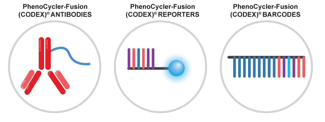
Three main components of PhenoCycler-Fusion (CODEX)®
Spatial biology multiplex imaging makes it possible to visualize in 3D and phenotype every cell across an entire microscope slide. By using DNA barcode technology comprised of unique oligonucleotide sequences conjugated to an antibody. PhenoCycler-Fusion can help to scale-up your spatial discovery efforts and translate those discoveries into actionable spatial phenotypic signatures.
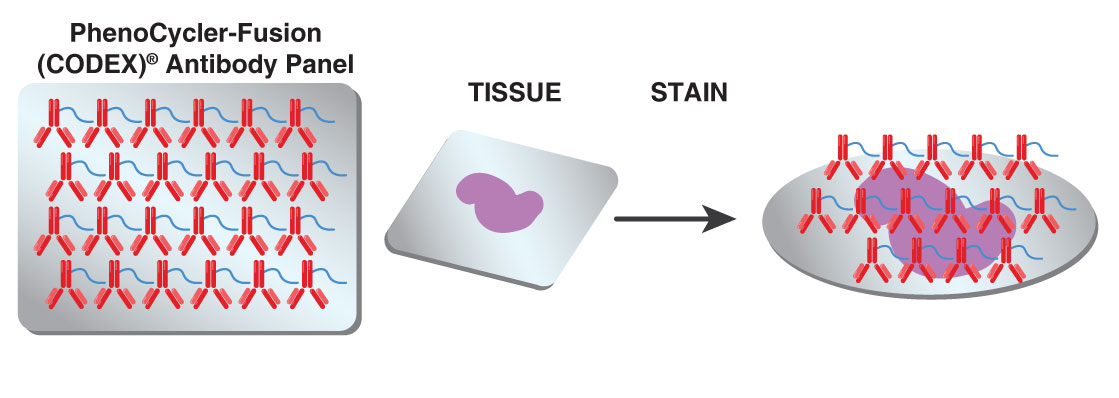
Single-step staining to preserve sample integrity
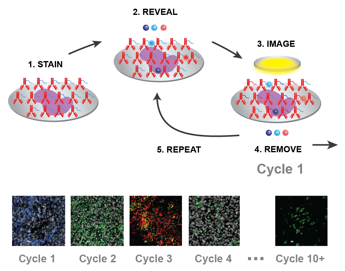
The PhenoCycler-Fusion (CODEX)® fluidics instrument has an automated imaging cycle. The first step is the staining of tissue with to >50 uniquely barcoded antibodies, combined for a single tissue staining reaction.
An automated process of imaging biomarkers using the PhenoCycler-Fusion (CODEX)® instrument and a companion microscope
Then, applied to the tissue and imaged are three reporter dyes with complimentary barcodes. Finally, the three reporter dyes are removed and the process is repeated until all antibodies have been imaged. Analyze 5 to 100+ slides per week.


