Fibroblast growth factor 10, also known as FGF10, is a member of the fibroblast growth factor (FGF) family. FGF family members possess broad mitogenic and cell survival activities, and are involved in a variety of biological processes, including embryonic development, cell growth, morphogenesis, tissue repair, tumor growth and invasion. This protein exhibits mitogenic activity for keratinizing epidermal cells, but essentially no activity for fibroblasts, which is similar to the biological activity of FGF7. FGF10 is a primary factor in the process of wound healing similarly to other growth factors such as TGF alpha and FGF7.1
Protein Details
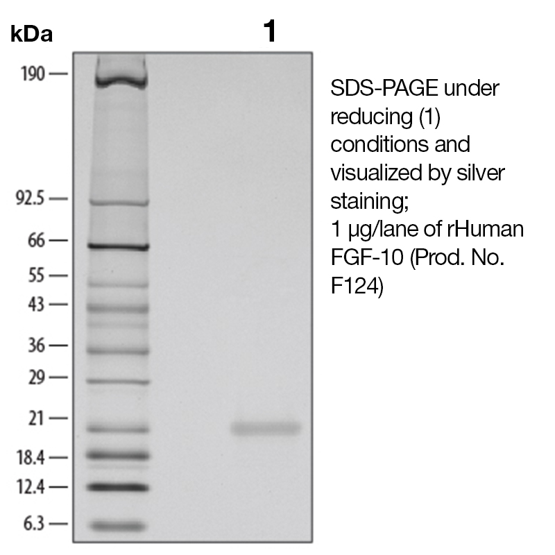
Purity
>97% by SDS-PAGE and analyzed by silver stain.
Endotoxin Level
<0.1 EU/µg as determined by the LAL method
Biological Activity
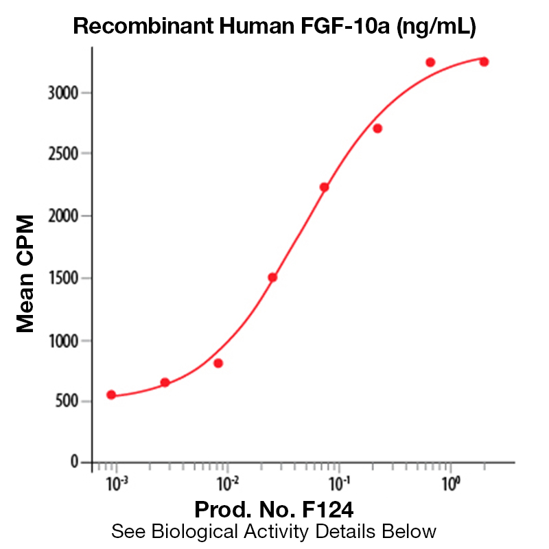
The biological activity of Human FGF-10 was determined by its ability to stimulate the proliferation of a monkey epithelial cell line, 4MBr-5 (Rubin, J.S. et al., 1989, Proc. Natl. Acad. Sci. USA 86:802). The expected ED<sub>50</sub> for this effect is typically 100 - 300 ng/ml.
The predicted molecular weight of Recombinant Human FGF-10 is Mr 19.5 kDa. However, the actual molecular weight as observed by migration on SDS-PAGE is Mr 19-22 kDa (reducing conditions).
Predicted Molecular Mass
19.5
Storage and Stability
This lyophilized protein is stable for six to twelve months when stored desiccated at -20°C to -70°C. After aseptic reconstitution, this protein may be stored at 2°C to 8°C for one month or at -20°C to -70°C in a manual defrost freezer. Avoid Repeated Freeze Thaw Cycles. See Product Insert for exact lot specific storage instructions.
Powered by AI: AI is experimental and still learning how to provide the best assistance. It may occasionally generate incorrect or incomplete responses. Please do not rely solely on its recommendations when making purchasing decisions or designing experiments.
Recombinant Human FGF-10 is widely used in research due to its critical roles in cell proliferation, differentiation, tissue development, and repair, making it valuable for studies in developmental biology, regenerative medicine, and disease modeling.
Key scientific reasons to use Recombinant Human FGF-10 in research applications:
Promotes Cell Proliferation and Survival: FGF-10 stimulates proliferation in various epithelial and mesenchymal cell types, supporting cell viability and expansion in vitro. This is particularly useful for culturing organoids, primary cells, and stem cells.
Essential for Developmental Biology: FGF-10 is a key regulator of embryonic development, including lung, limb, kidney, brain, and heart morphogenesis. It is indispensable for modeling organogenesis and studying developmental pathways.
Supports Organoid and Tissue Engineering: FGF-10 is commonly used to drive the growth and maintenance of organoids (e.g., lung, liver, salivary gland), enabling the creation of physiologically relevant in vitro models for disease research and drug testing.
Facilitates Stem Cell Differentiation: It is critical for the differentiation of pluripotent stem cells (ES/iPS cells) into specific lineages, especially those of endodermal and mesodermal origin, such as lung and glandular tissues.
Tissue Repair and Regeneration: FGF-10 has demonstrated roles in wound healing, epithelial repair, and protection against tissue injury, including in the lung and skin. It is used to model and study regenerative processes and tissue responses to injury.
Disease Modeling: FGF-10 is involved in the pathogenesis and repair mechanisms of diseases such as chronic obstructive pulmonary disease (COPD), fibrosis, and hormone-dependent cancers. Its use enables the study of disease mechanisms and therapeutic interventions.
Enhanced Stability for Consistent Results: Engineered, heat-stable forms of recombinant FGF-10 are available, offering greater thermal stability and prolonged activity in cell culture, which improves reproducibility and efficacy in long-term experiments.
Paracrine Signaling Studies: FGF-10 acts as a paracrine growth factor, making it valuable for dissecting cell–cell communication and niche interactions in both development and disease contexts.
Typical applications include:
Cell proliferation and viability assays
Directed differentiation of stem cells
Organoid culture and tissue engineering
Wound healing and tissue repair models
Developmental biology and morphogenesis studies
Disease modeling and drug screening
In summary, Recombinant Human FGF-10 is a versatile and essential tool for research focused on cell growth, tissue development, regeneration, and disease modeling, with broad applications across basic and translational biomedical sciences.
Yes, recombinant human FGF-10 can be used as a standard for quantification or calibration in ELISA assays, provided it is compatible with your assay system and matches the form used by the kit manufacturer. Several ELISA kits for FGF-10 quantification use recombinant human FGF-10 as their standard, and suppliers specifically recommend recombinant FGF-10 (often with BSA as a carrier) for use as an ELISA standard.
Key considerations for use:
Formulation: Recombinant FGF-10 with BSA is generally recommended for use as an ELISA standard, as BSA helps stabilize the protein and prevent adsorption to plasticware. Carrier-free formulations are available but may be less stable and more prone to loss during handling.
Source and Sequence: Ensure the recombinant FGF-10 matches the sequence and post-translational modifications (if any) expected by your ELISA kit. Most kits and standards use E. coli-expressed FGF-10, which is suitable for most immunoassays.
Validation: The recombinant standard should be validated for use in your specific ELISA system. Some kits specify that their calibration curve is generated using recombinant FGF-10, and parallelism between recombinant and native FGF-10 is often demonstrated.
Concentration and Reconstitution: Follow the manufacturer’s instructions for reconstitution and dilution to ensure accurate standard curve generation.
Limitations and best practices:
Matrix Effects: If you are quantifying FGF-10 in complex biological samples (e.g., serum, plasma), matrix effects may influence recovery and quantification. It is best practice to prepare the standard curve in the same matrix as your samples, or to use appropriate diluents recommended by the kit.
Kit Compatibility: Always check your ELISA kit’s documentation to confirm that recombinant FGF-10 is suitable as a standard. Some kits may require a specific isoform or formulation for optimal performance.
Bioactivity vs. Immunoreactivity: ELISA standards require immunoreactivity (recognition by the capture and detection antibodies), not necessarily bioactivity. However, most commercial recombinant FGF-10 proteins are validated for both.
Summary Table: Use of Recombinant FGF-10 as ELISA Standard
Requirement
Recommendation/Note
Protein source
Recombinant human FGF-10 (E. coli-expressed common)
Formulation
With BSA carrier preferred for ELISA standard
Validation
Confirm with kit documentation
Matrix for standard curve
Match to sample matrix if possible
Reconstitution
Follow supplier/kit instructions
Bioactivity needed?
No, only immunoreactivity is required
In conclusion, recombinant human FGF-10 is widely accepted and recommended as a standard for ELISA quantification, provided it is compatible with your assay system and prepared according to best practices.
Recombinant Human FGF-10 has been validated for a broad range of applications in published research, particularly in studies involving cell signaling, development, tissue repair, and disease modeling.
Key validated applications include:
Cell culture and functional assays: FGF-10 is widely used to stimulate proliferation and differentiation of epithelial cells, including in studies of lung, limb, and glandular development.
Stem cell differentiation: It is used to direct the differentiation of embryonic stem (ES) and induced pluripotent stem (iPS) cells, especially toward epithelial and mesenchymal lineages relevant to lung and organoid models.
Organoid culture: FGF-10 supports the growth and maintenance of organoids derived from various tissues, such as liver, lung, and salivary gland, and is used in bioassays to model tissue development and disease.
Bioassays and proliferation studies: It is validated for use in bioactivity assays, including proliferation of human urothelial and transitional epithelial cells.
ELISA and Western blot: FGF-10 is used as a standard or control in ELISA and Western blot assays to quantify or detect FGF-10 or its downstream signaling components.
Immunohistochemistry: It is used to study tissue localization and expression patterns of FGF-10 in developmental and disease contexts.
In vivo models: FGF-10 has been used in animal models to study its role in lung injury, repair, and inflammation, including mobilization of lung-resident mesenchymal stem cells (LR-MSCs) and attenuation of acute lung injury.
Cancer research: Its role in carcinogenesis and epithelial-mesenchymal interactions has been explored in models of prostate, breast, pancreas, and colorectal cancer.
Wound healing: FGF-10 is applied in studies of epithelial repair and regeneration in both normal and wounded tissues.
References to specific published research confirm these applications, including studies on lung organoid development, acute lung injury models, and stem cell differentiation protocols.
If you need protocol details or specific references for a particular application, please specify the context or research area.
To reconstitute and prepare Recombinant Human FGF-10 protein for cell culture experiments, first briefly centrifuge the vial to collect the lyophilized powder at the bottom. Reconstitute the protein in either sterile distilled water or sterile PBS (phosphate-buffered saline), depending on the formulation and downstream application. For optimal stability and activity in cell culture, it is recommended to include a carrier protein such as 0.1–1% bovine serum albumin (BSA) or human serum albumin (HSA) in the buffer.
Step-by-step protocol:
Centrifuge the vial briefly before opening to ensure all powder is at the bottom.
Reconstitution concentration: Prepare a stock solution at 100 μg/mL in sterile PBS containing 0.1% BSA (or HSA) for most cell culture applications. Some protocols allow concentrations up to 0.1–1.0 mg/mL in sterile water or buffer.
Gentle handling: Avoid vigorous pipetting or vortexing to prevent protein denaturation.
Aliquot the stock solution to minimize freeze-thaw cycles.
Storage: Store reconstituted protein at 4°C for up to 1 month, or at –20°C to –80°C for longer periods. Avoid repeated freeze-thaw cycles to maintain bioactivity.
Working solution: Dilute the stock solution to the desired final concentration in cell culture medium immediately before use. Typical working concentrations for cell proliferation assays range from 20–100 ng/mL.
Additional notes:
If the protein is supplied carrier-free, reconstitute in sterile PBS or water and add carrier protein (BSA/HSA) to prevent adsorption and loss of activity, especially at low concentrations.
For organoid or stem cell differentiation protocols, follow specific medium recipes and supplement with FGF-10 at the recommended concentration.
If long-term storage is required, consider adding 5–50% glycerol to the stock solution for enhanced stability.
Summary Table:
Step
Buffer/Condition
Concentration
Additive
Notes
Centrifuge vial
—
—
—
Collect powder at bottom
Reconstitute
Sterile PBS or water
100 μg/mL (typical)
0.1–1% BSA/HSA
Gentle pipetting, no vortex
Aliquot
—
—
—
Avoid freeze-thaw cycles
Store
4°C or –20°C to –80°C
—
—
Use within 1–3 months
Working dilution
Cell culture medium
20–100 ng/mL
—
Prepare fresh before use
Always consult the specific product datasheet for formulation details and recommended diluents, as requirements may vary depending on the recombinant protein’s source and intended application.


 Products are for research use only. Not for use in diagnostic or therapeutic procedures.