Fibroblast growth factor receptor-1 beta (FGFR1- beta) is an isoform of FGFR1 which is a receptor tyrosine kinase. FGFR1-beta is expressed in basal epithelial cells and smooth muscle cells.1 FGF R1β supports progression and chemoresistance in subsets of acute myeloid leukemias (AML).2
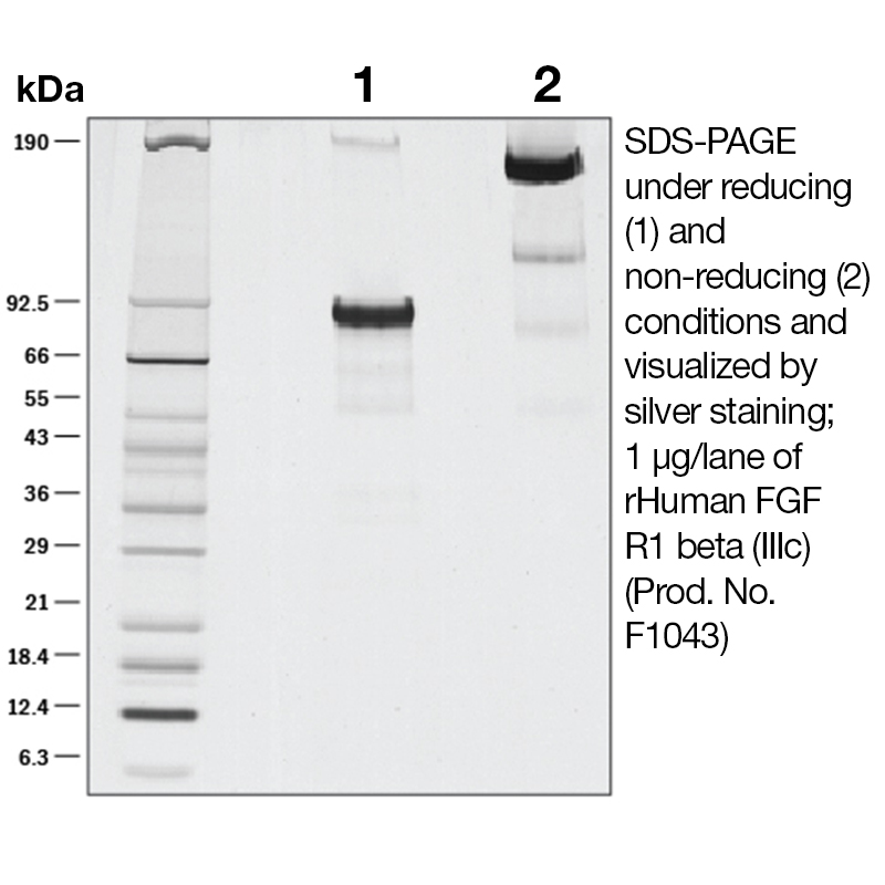
The predicted molecular weight of Recombinant Human FGF R1β (IIIc) is Mr 56 kDa. However, the actual molecular weight as observed by migration on SDS-PAGE is Mr 90-95 kDa.
Predicted Molecular Mass
56
Formulation
This recombinant protein was 0.2 µm filtered and lyophilized from modified Dulbecco’s phosphate buffered saline (1X PBS) pH 7.2 – 7.3 with no calcium, magnesium, or preservatives.
Storage and Stability
This lyophilized protein is stable for six to twelve months when stored desiccated at -20°C to -70°C. After aseptic reconstitution, this protein may be stored at 2°C to 8°C for one month or at -20°C to -70°C in a manual defrost freezer. Avoid Repeated Freeze Thaw Cycles. See Product Insert for exact lot specific storage instructions.
Powered by AI: AI is experimental and still learning how to provide the best assistance. It may occasionally generate incorrect or incomplete responses. Please do not rely solely on its recommendations when making purchasing decisions or designing experiments.
Recombinant Human FGF R1β (IIIc) is a specialized isoform of the fibroblast growth factor receptor 1 (FGFR1), a receptor tyrosine kinase involved in key cellular signaling pathways. Using this recombinant protein in research applications enables precise investigation of FGF signaling, cellular responses, and disease mechanisms, particularly those involving epithelial and smooth muscle cells.
Key scientific reasons to use Recombinant Human FGF R1β (IIIc):
Isoform specificity: The IIIc isoform of FGFR1β is distinct in its ligand-binding properties and tissue distribution, allowing researchers to dissect the specific roles of FGF signaling in different cell types and physiological contexts.
Functional studies: Recombinant FGF R1β (IIIc) is essential for studying FGF-dependent processes such as cell proliferation, migration, differentiation, and survival, which are relevant in development, tissue repair, and cancer biology.
Disease modeling: FGFR1β (IIIc) supports progression and chemoresistance in certain acute myeloid leukemia (AML) subsets, making it valuable for cancer research and drug screening.
Assay development: This protein is used in binding assays, ELISA, and cell-based functional assays to characterize FGF ligand-receptor interactions and downstream signaling events.
Receptor-ligand specificity: FGF-21 and other FGFs show selective signaling through FGFR1 isoforms, including IIIc, which is crucial for understanding tissue-specific responses and developing targeted therapeutics.
Technical considerations:
High purity and stability: Recombinant preparations are typically >90% pure and endotoxin-free, ensuring reliable results in sensitive assays and cell culture experiments.
Storage and handling: Lyophilized recombinant FGF R1β (IIIc) is stable for extended periods when stored properly, minimizing degradation and variability in experimental outcomes.
Interference in multiplex assays: Recombinant FGF R1β (IIIc) may interfere with certain immunoassays at high concentrations, so appropriate controls and optimization are necessary for quantitative studies.
Applications include:
Cell signaling and receptor activation studies
Cancer biology and chemoresistance research
Tissue engineering and regenerative medicine
Development of diagnostic and therapeutic assays
In summary, Recombinant Human FGF R1β (IIIc) is a critical tool for dissecting FGF signaling pathways, modeling disease mechanisms, and developing targeted interventions in biomedical research.
Recombinant Human FGF R1β (IIIc) can be used as a standard for quantification or calibration in ELISA assays, provided it is highly purified, its concentration is accurately determined, and it matches the analyte detected by your assay. However, several technical considerations must be addressed to ensure accurate quantification:
Purity and Characterization: The recombinant FGF R1β (IIIc) you referenced is reported as >90% pure by SDS-PAGE and silver stain, with low endotoxin levels and a defined molecular mass. High purity and accurate quantification of the standard are essential for reliable ELISA calibration.
Standard Curve Preparation: For quantitative ELISA, a standard curve must be generated using known concentrations of the standard protein. The recombinant protein should be reconstituted and diluted according to best practices, ensuring consistency and accuracy in concentration.
Isoform and Epitope Matching: The standard must be the same isoform and have the same epitopes as the analyte detected by your ELISA antibodies. FGF R1β (IIIc) is a specific isoform; if your ELISA is designed to detect this isoform, it is appropriate as a standard. If your assay detects a different isoform or domain, using this standard may result in inaccurate quantification due to differences in antibody recognition.
Cross-reactivity and Interference: Some ELISA kits report cross-reactivity or interference from closely related proteins or isoforms. Ensure that your assay is validated for FGF R1β (IIIc) and that the recombinant standard does not introduce unexpected cross-reactivity.
Validation: It is best practice to validate the use of any new standard by comparing results with a previously validated standard curve or by spike-and-recovery experiments to confirm accuracy and linearity.
Summary of best practices:
Use a highly purified, well-characterized recombinant protein as your standard.
Confirm that the standard matches the analyte detected by your ELISA antibodies (same isoform and relevant epitopes).
Prepare the standard curve carefully, following established protocols for dilution and reconstitution.
Validate the standard in your assay system to ensure accurate quantification.
If your ELISA is specifically designed for FGF R1β (IIIc), and the recombinant protein is of high quality and correctly quantified, it is suitable for use as a standard. If your assay targets a different isoform or domain, select a standard that matches your analyte for optimal accuracy.
Recombinant Human FGF R1β (IIIc) has been validated in published research primarily for applications involving biochemical assays, receptor-ligand interaction studies, and multiplex cytokine detection platforms.
Key validated applications include:
Multiplex Cytokine Assays: Recombinant Human FGF R1β (IIIc) is used as a standard or analyte in multiplex immunoassays, such as Luminex®-based platforms, for the quantitative detection of FGF R1β (IIIc) in biological samples. These assays are widely used in research to profile cytokine and growth factor levels in various experimental contexts, including disease models and clinical samples.
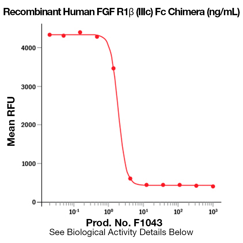
Receptor-Ligand Binding Studies: The recombinant protein, particularly when fused to an Fc domain, is used in biochemical and cell-based assays to study the binding specificity and affinity of FGF ligands (such as FGF2) to the FGFR1β (IIIc) isoform. This is essential for dissecting FGF signaling pathways and for screening potential therapeutic inhibitors or agonists.
Cancer Research: FGFR1β (IIIc) has been implicated in supporting progression and chemoresistance in subsets of acute myeloid leukemia (AML), and recombinant forms are used in functional studies to elucidate these mechanisms. Such studies may involve cell proliferation, survival, and drug resistance assays in vitro.
Protein Characterization and Quality Control: The recombinant protein is validated by SDS-PAGE, silver staining, and endotoxin testing for use in research applications requiring high purity and low endotoxin levels.
Additional context:
The recombinant protein is typically produced as an Fc-fusion, which facilitates purification and detection in various assay formats.
While the search results do not report direct in vivo therapeutic studies with FGF R1β (IIIc) itself, its use in multiplex assays and receptor studies is well established in the literature.
If you require protocols or best practices for a specific application (e.g., ELISA, flow cytometry, cell signaling assays), please specify the intended use.
To reconstitute and prepare Recombinant Human FGF R1β (IIIc) protein for cell culture experiments, follow these steps to ensure protein stability and biological activity:
Centrifuge the vial briefly before opening to collect all lyophilized powder at the bottom.
Reconstitute the protein using sterile buffer. For FGF R1β (IIIc) lyophilized from PBS (pH 7.2–7.3, no Ca²⁺/Mg²⁺), use sterile PBS or a similar physiological buffer. If the product was lyophilized from a different buffer (e.g., Tris), match the buffer for reconstitution.
Recommended concentration: Prepare a stock solution at 0.1–1.0 mg/mL, depending on your experimental needs. For example, add 100–1000 µL buffer per 0.1 mg protein.
Mix gently to dissolve the protein, avoiding vigorous agitation which may denature it.
Aliquot the solution to minimize freeze-thaw cycles, which can reduce activity.
Storage after reconstitution: Store aliquots at 2–8°C for up to one month, or at –20°C to –70°C for longer periods. Avoid repeated freeze-thaw cycles.
For cell culture: Dilute the stock solution in cell culture medium immediately before use. Include a carrier protein (e.g., 0.1% BSA) in working solutions to stabilize the protein and prevent adsorption to plasticware.
Additional notes:
Confirm the absence of endotoxin if using in sensitive cell types; typical preparations have <0.01 EU/µg endotoxin.
Always consult the specific product datasheet for lot-specific instructions, as formulation and recommended buffers may vary.
For functional assays, typical working concentrations range from low ng/mL to several hundred ng/mL, depending on cell type and experimental design.
Summary protocol:
Centrifuge vial, open aseptically.
Add sterile PBS (or specified buffer) to achieve 0.1–1.0 mg/mL.
Mix gently until fully dissolved.
Aliquot and store at –20°C to –70°C.
Dilute in cell culture medium with carrier protein for experiments.
This protocol ensures optimal recovery, stability, and activity of recombinant FGF R1β (IIIc) for cell culture applications.
References & Citations
1. Moscatelli, D. et al. (2007) Prostate67: 115
2. Raffi, S. et al. (2006) Leukemia20: 979


 Products are for research use only. Not for use in diagnostic or therapeutic procedures.