

Anti-LRP4 (extracellular) Antibody (56540)


Anti-LRP4 (extracellular) Antibody (56540)
Product No.: 56540
- -
- -
Clone S207-27 Target LRP4 (extracellular) Formats AvailableView All Product Type Monoclonal Alternate Names LRP-4, LDLR dan Isotype Mouse IgG2a Applications ICC , IF , WB |
Data
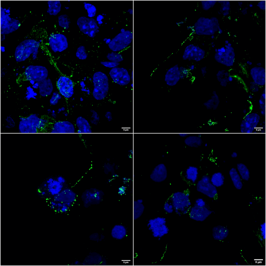 Immunocytochemistry/Immunofluorescence analysis using Mouse Anti-LRP4 Monoclonal Antibody, Clone S207-27 (56540). Tissue: COS cells transfected with 2 ug human LRP4 plasmid. Species: Human. Fixation: 4% PFA for 10 min. Primary Antibody: Mouse Anti-LRP4 Monoclonal Antibody (56540) at 1:100 for 1 hour at RT. Secondary Antibody: Goat anti-mouse: Alexa 488. Counterstain: DAPI.
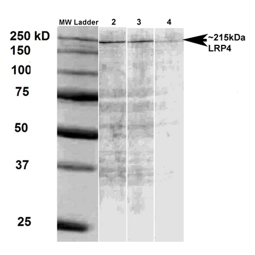 Western Blot analysis of Rat brain membrane lysate showing detection of LRP4 protein using Mouse Anti-LRP4 Monoclonal Antibody, Clone S207-27 (56540). Primary Antibody: Mouse Anti-LRP4 Monoclonal Antibody (56540) at 1:500, 1:1000, and 1:2000.
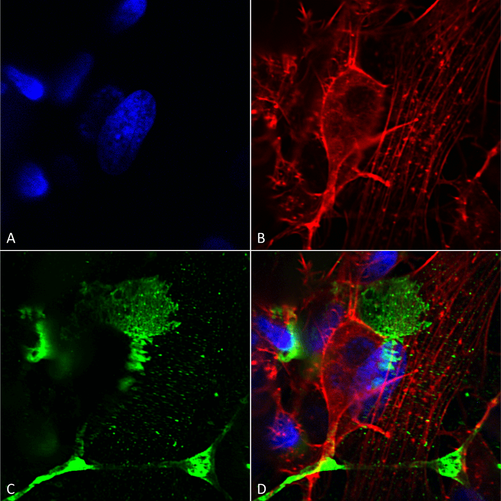 Immunocytochemistry/Immunofluorescence analysis using Mouse Anti-LRP4 (Extracellular) Monoclonal Antibody, Clone S207-27 (56540). Tissue: Neuroblastoma cells (SH-SY5Y). Species: Human. Fixation: 4% PFA for 15 min. Primary Antibody: Mouse Anti-LRP4 (Extracellular) Monoclonal Antibody (56540) at 1:200 for overnight at 4°C with slow rocking. Secondary Antibody: AlexaFluor 488 at 1:1000 for 1 hour at RT. Counterstain: Phalloidin-iFluor 647 (red) F-Actin stain; Hoechst (blue) nuclear stain at 1:800, 1.6mM for 20 min at RT. (A) Hoechst (blue) nuclear stain. (B) Phalloidin-iFluor 647 (red) F-Actin stain. (C) LRP4 (Extracellular) Antibody (D) Composite.
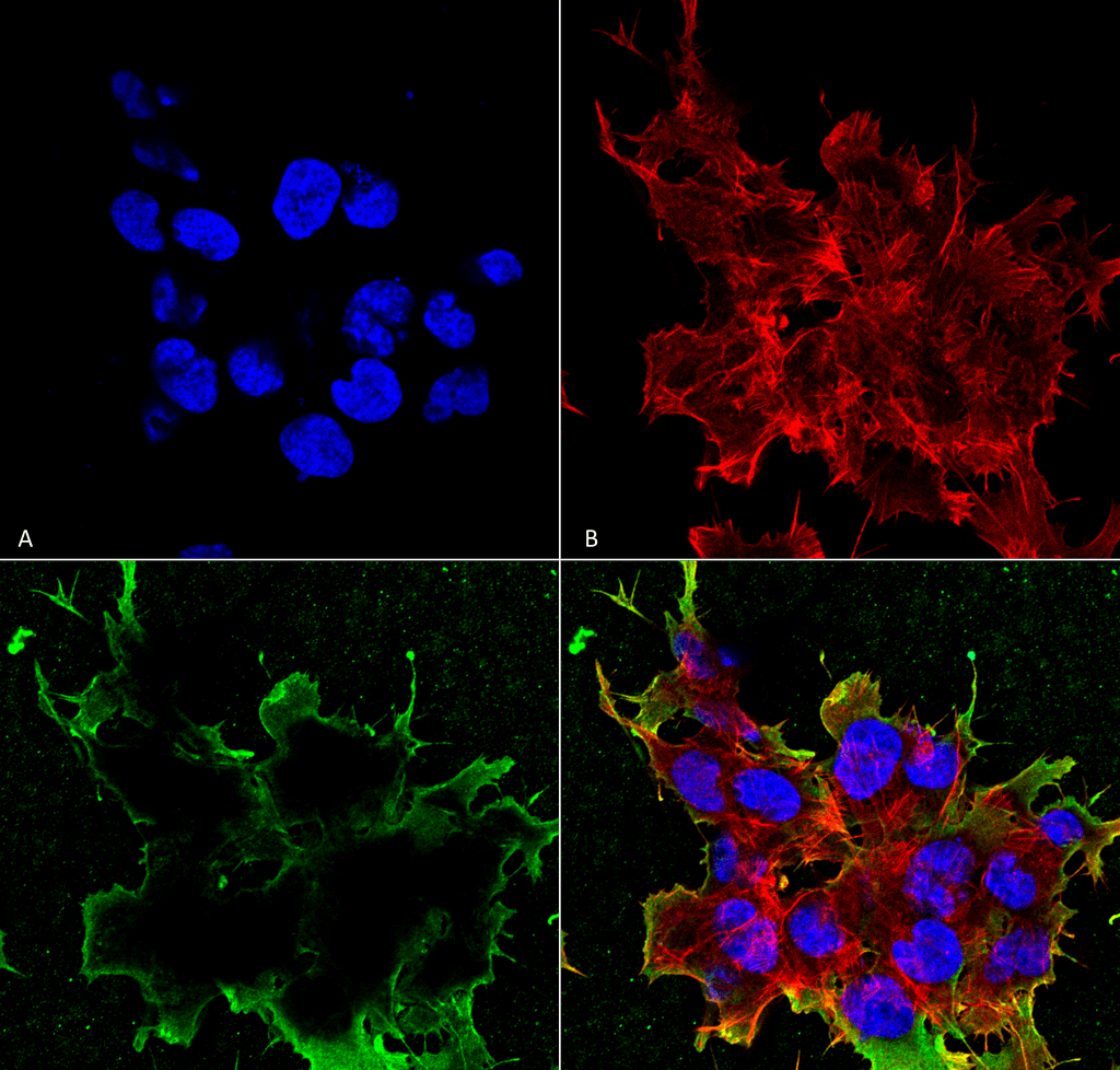 Immunocytochemistry/Immunofluorescence analysis using Mouse Anti-LRP4 (Extracellular) Monoclonal Antibody, Clone S207-27 (56540). Tissue: Neuroblastoma cell line (SK-N-BE). Species: Human. Fixation: 4% Formaldehyde for 15 min at RT. Primary Antibody: Mouse Anti-LRP4 (Extracellular) Monoclonal Antibody (56540) at 1:100 for 60 min at RT. Secondary Antibody: Goat Anti-Mouse ATTO 488 at 1:100 for 60 min at RT. Counterstain: Phalloidin Texas Red F-Actin stain; DAPI (blue) nuclear stain at 1:1000, 1:5000 for 60min RT, 5min RT. Localization: Membrane. Magnification: 60X. (A) DAPI (blue) nuclear stain. (B) Phalloidin Texas Red F-Actin stain. (C) LRP4 (Extracellular) Antibody. (D) Composite.
 - -
- -
Antibody DetailsProduct DetailsReactive Species Mouse ⋅ Rat Host Species Mouse Immunogen Fusion protein corresponding to aa 26-350 (extracellular N-terminus) of mouse LRP4 (accession no.Q8V156). Product Concentration 1.0 mg/ml Formulation PBS, pH 7.4, 50% glycerol, 0.09% sodium azide.Purified by Protein G affinity chromatography. State of Matter Liquid Product Preparation Purified by Protein G affinity chromatography Storage and Handling This antibody is stable for at least one (1) year at -20°C. Avoid repeated freeze-thaw cycles. Regulatory Status For in vitro investigational use only. Not
intended for diagnostic or therapeutic
applications. Country of Origin USA Shipping Next Day 2-8°C Applications and Recommended Usage? Quality Tested by Leinco Immunoblotting: use at 1ug/mL. Predicted molecular weight is ~215kDa. Degradation products of approx. 150kDa are also detected.
Positive control: rat brain lysate. These are recommended concentrations. Endusers should determine optimal concentrations for their applications. Each investigator should determine their own optimal working dilution for specific applications. See directions on lot specific datasheets, as information may periodically change. DescriptionDescriptionSpecificity This antibody recognizes mouse and rat
LRP4. Background The formation of neuromuscular junctions (NMJ) requires the interaction between motor neurons and muscle fibers. LRP4 (low-density lipoprotein receptor-related protein 4) is a receptor of agrin that is thought to act in cis to stimulate MuSK in muscle fibers for postsynaptic differentiation. Recent studies of muscle- specific mutants suggest that LRP4 is involved in determining where AChR clusters form in muscle fibers, postsynaptic differentiation, and axon terminal development. Function Mediates SOST-dependent inhibition of bone formation. Functions as a specific facilitator of SOST-mediated inhibition of Wnt signaling. Plays a key role in the formation and the maintenance of the neuromuscular junction (NMJ), the synapse between motor neuron and skeletal muscle. Directly binds AGRIN and recruits it to the MUSK signaling complex. Mediates the AGRIN-induced phosphorylation of MUSK, the kinase of the complex. The activation of MUSK in myotubes induces the formation of NMJ by regulating different processes including the transcription of specific genes and the clustering of AChR in the postsynaptic membrane. Alternatively, may be involved in the negative regulation of the canonical Wnt signaling pathway, being able to antagonize the LRP6-mediated activation of this pathway. More generally, has been proposed to function as a cell surface endocytic receptor binding and internalizing extracellular ligands for degradation by lysosomes. Plays an essential role in the process of digit differentiation (PubMed:16517118). {PubMed:16517118, PubMed:18848351, PubMed:21471202}. NCBI Gene Bank ID UniProt.org Research Area Neuroscience References & CitationsTechnical ProtocolsICC IF  Certificate of Analysis |
Formats Available
 Products are for research use only. Not for use in diagnostic or therapeutic procedures.