

Anti-Mouse CD223 (LAG-3) [C9B7W] ; Alexa Fluor™ 647-RX033

Anti-Mouse CD223 (LAG-3) [C9B7W] ; Alexa Fluor™ 647-RX033
Product No.: L310
- -
- -
Clone C9B7W Target LAG-3 CD223 Formats AvailableView All Product Type Monoclonal Antibody Alternate Names CD223, LAG3 Isotype Rat IgG1 κ Applications IHC FF , PhenoCycler® |
Data
- -
- -
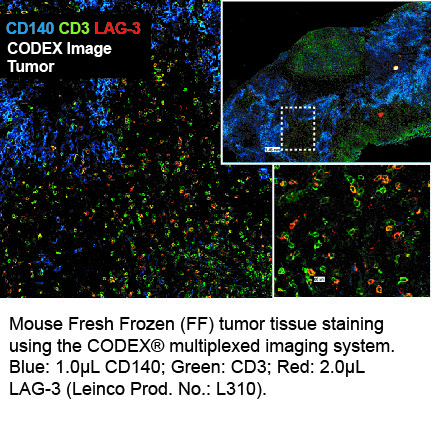
CODEX® DetailsBarcode BX033 Reporter Alexa Fluor™ 647-RX033 Tissue Screened MCA Sarcoma (Tumor) Tissue Preparation FF (Fresh Frozen) Antibody and Reporter DetailsReactivity Species Mouse Host Species Rat Immunogen Murine CD223-Ig fusion protein Formulation This PhenoCycler-Fusion (CODEX)® barcoded antibody is formulated in phosphate buffered saline (150 mM NaCl) PBS, EDTA pH 7.2 containing 0.09% sodium azide as a preservative. The CODEX® reporter is formulated in 1X Tris-EDTA (TE) pH 8.0 (10 mM Tris-HCl, 1 mM disodium EDTA, pH 8.0) Product Preparation Manufactured in an animal-free facility using only in vitro cell culture techniques, purified by affinity chromatography, and conjugated to a specific barcode under optimal conditions. State of Matter CODEX® barcoded antibody - Liquid; CODEX® reporter - Liquid Storage and Handling This PhenoCycler-Fusion (CODEX)® barcoded antibody is stable when stored at 2-8°C for up to 1 year (do not freeze). The CODEX® reporter is stable when frozen at -20°C for up to 1 year. Applications and Recommended Usage? Quality Tested by Leinco CODEX®; IHC FF - The suggested dilution for staining tissue in immunohistochemistry - Fresh Frozen (FF) is 1 μl of Anti-Mouse LAG-3 (BX033) in a final volume of 200 μl of CODEX® staining buffer. Country of Origin USA Each investigator should determine their own optimal working dilution for specific applications. See directions on lot specific datasheets, as information may periodically change. DescriptionSpecificity Clone C9B7W recognizes and specifically binds to an epitope in the D2 domain of CD223. Antigen Distribution CD223 is expressed on T regulatory cells, activated T cells and NK cells. Background Antibody LAG-3 is a 70-kD, type-I transmembrane glycoprotein within the Ig superfamily with four extracellular Ig-like domains (D1 to D4) and is structurally homologous to CD4. LAG-3 is a cell surface molecule with various biologic effects on T cell function. It has been reported to be involved in Treg suppressive function. It negatively regulates cellular proliferation, activation, and homeostasis of T cells, in a similar manner to CTLA-4 and PD-1. Human LAG-3 is approximately 70% homologous with murine LAG3, and it binds MHC class II molecules with higher affinity than CD4. As an immune checkpoint receptor, LAG-3 is the target of various drug development programs seeking to expand treatments for cancer and autoimmune disorders. In its soluble form, LAG-3 is being developed as a cancer drug. As an antagonist, LAG-3 antibody can activate T effector cells via the downregulation of the LAG-3 inhibiting signal into pre-activated LAG-3+ cells. In addition, it can inhibit antigen-specific Treg suppressive activity. As an agonist antibody, it can be used to diminish an autoimmune response and is currently being investigated for the treatment of plaque psoriasis.
PhenoCycler-Fusion (CODEX)® reporters are made up of a fluorescent dye and a short oligonucleotide DNA barcode called a CODEX® Tag. Fluorescent reporters enable highly specific detection of corresponding barcodes, and the use of spectrally separated dyes allow for precise signal detection in up to three distinct fluorescence channels at one time. Antigen DetailsProtein PubMed NCBI Gene Bank ID Technical Protocols PhenoCycler® |
Formats Available
- -
- -
Prod No. | Description |
|---|---|
C2252 | |
C2253 | |
C2254 | |
C2255 | |
C2256 | |
C2249 | |
C2251 | |
C2250 | |
L306 | |
C2155 | |
L502 | |
C2852 | |
L313 | |
L310 |
