

Anti-Mouse Siglec-F-BX016; Alexa Fluor™ 488 – RX016

Anti-Mouse Siglec-F-BX016; Alexa Fluor™ 488 – RX016
Product No.: C2878
- -
- -
Clone E50-2440 Target Siglec-F Formats AvailableView All Product Type Monoclonal Antibody Isotype Mouse IgG2a k Applications ELISA , FC , IF Microscopy , IHC , IHC FF , PhenoCycler® |
Data
- -
- -
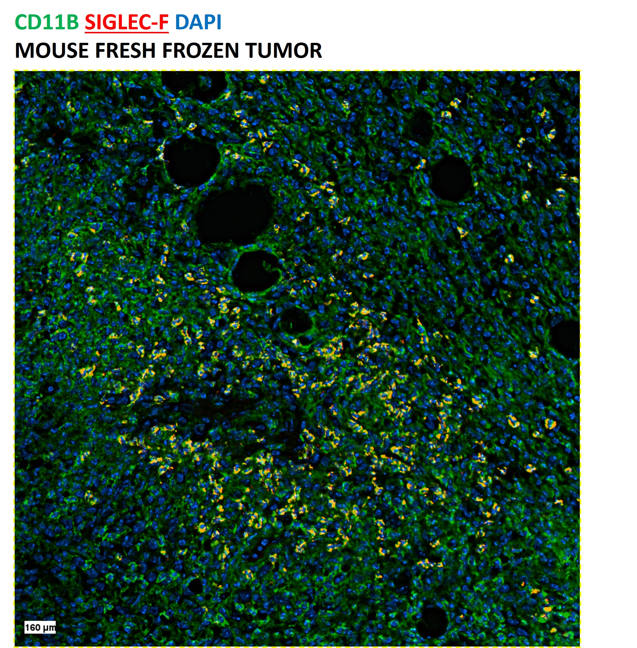
CODEX® DetailsBarcode BX016 Reporter Alexa Fluor™ 488-RX016 Fluorescent Dye Alexa Fluor™ 488 is in the bright green fluorescent channel with an excitation max of 490 nm and an emission max of 525 nm. Tissue Screened Tonsil Tissue Preparation FF (Fresh Frozen) Antibody and Reporter DetailsReactivity Species Rat Host Species Mouse Formulation This PhenoCycler-Fusion (CODEX)® barcoded antibody is formulated in phosphate buffered saline (150 mM NaCl) PBS, EDTA pH 7.2 containing 0.09% sodium azide as a preservative. The CODEX® reporter is formulated in 1X Tris-EDTA (TE) pH 8.0 (10 mM Tris-HCl, 1 mM disodium EDTA, pH 8.0) Product Preparation Manufactured in an animal-free facility using only in vitro cell culture techniques, purified by affinity chromatography, and conjugated to a specific barcode under optimal conditions. State of Matter CODEX® barcoded antibody - Liquid; CODEX® reporter - Liquid Storage and Handling This PhenoCycler-Fusion (CODEX)® barcoded antibody is stable when stored at 2-8°C for up to 1 year (do not freeze). The CODEX® reporter is stable when frozen at -20°C for up to 1 year. Applications and Recommended Usage? Quality Tested by Leinco CODEX®; IHC FF - The suggested dilution for staining tissue in immunohistochemistry - Fresh Frozen (FF) is 1-2 μl of Anti-Mouse CD140A (BX016) in a final volume of 200 μl of CODEX® staining buffer.
ELISA4, FC3, 4, 5, IHC4, IF Microscopy4, B3, Country of Origin USA Each investigator should determine their own optimal working dilution for specific applications. See directions on lot specific datasheets, as information may periodically change. DescriptionSpecificity E50-2440 activity is directed against mouse Siglec-F. Antigen Distribution Siglec-F positive cells are expressed by eosinophils, alveolar macrophages, thymus and lung parenchyma tissues, and at very low levels on T cells. Siglec-F expression is restricted to cells of myelomonocytic lineage (CD11b-positive). Siglec-F is present on CD11blo (lo = low expressors) and CD11bhi (hi = low expressors) cell populations from mouse bone marrow, blood, and spleen but is especially high on the CD11blo cells. Cells that are CD11blo/Siglec-Fhi are predominantly immature cells of the myelomonocytic lineage, and cells that are CD11blo-hi/Siglec-Flo are mostly mature or immature neutrophils and monocytes, including leukocytes. Siglec-F is not expressed on mouse mast cells, stem cells, B cells, or NK cells. Background Siglecs (sialic acid-binding immunoglobulin superfamily lectins) are a family of single pass, transmembrane cell surface proteins characterized by shared structural motifs and an ability to recognize sialic acids1,2. Siglec-F selectively recognizes 6'-sulfo-sialyl Lewis X as a glycan ligand, and this interaction is inhibited by the E50-2440 antibody3. Siglec-F belongs to the Siglec-3 (CD33) group4 and is a functionally convergent paralog of human Siglec-83. Mouse Siglec-F is used as a model to understand glycan based therapeutic targeting strategies in humans1, 2.
Expression of Siglec-F on eosinophils3 plays a role in the allergic inflammatory response, during which its surface levels on eosinophils and other cells increase1. Siglec-F deficient mice that are exposed to allergen sensitization and asthma-inducing conditions display more pronounced bone marrow, blood, and tissue eosinophilia due to reduced apoptosis1 as well as exaggerated eosinophilic inflammation and tissue remodeling in asthma models2. Administration of Siglec-F antibody reduces blood and tissue eosinophils in vivo2. The rat monoclonal antibody E50-2440 was prepared by immunizing Lou rats with mSiglec-F-Fc, a fusion protein of the first two Ig-like domains of mouse Siglec-F and human IgG Fc fragment separated by an enterokinase cleavage site/FLAG tag (DYKDDDDK)4. Spleen cells were fused with Yb/20 myeloma cells, and hybridomas were screened by ELISA, tested by flow cytometry, and isotyped. The E50-2440 clone was selected based on its high staining intensity on primary cells. Siglec-F is a reliable marker of eosinophils among granulocytes in mouse1, and cellular staining profiles are identical in the C3H, Balb/c, and C57BL/6 mouse strains4. Antigen DetailsProtein PubMed References & Citations1. Bochner BS. Clin Exp Allergy. 39(3):317-324. 2009.
2. Kiwamoto T, Kawasaki N, Paulson JC, et al. Pharmacol Ther. 135(3):327-336. 2012. 3. Tateno H, Crocker PR, Paulson JC. Glycobiology. 15(11):1125-1135. 2005. 4. Angata T, Hingorani R, Varki NM, et al. J Biol Chem. 276(48):45128-45136. 2001. 5. Zhang JQ, Biedermann B, Nitschke L, et al. Eur J Immunol. 34(4):1175-1184. 2004. Technical Protocols  IF Microscopy  PhenoCycler® |
Formats Available
- -
- -
