

Anti-PINK1 Antibody (56548)


Anti-PINK1 Antibody (56548)
Product No.: 56548
- -
- -
Clone S4-15 Target PINK1 Formats AvailableView All Product Type Monoclonal Isotype Mouse IgG1 Applications ICC , IF , IHC , WB |
Data
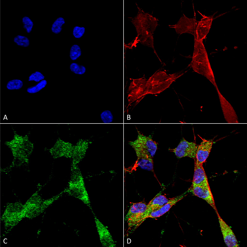 Immunocytochemistry/Immunofluorescence analysis using Mouse Anti-PINK1 Monoclonal Antibody, Clone S4-15 (56548). Tissue: Neuroblastoma cells (SH-SY5Y). Species: Human. Fixation: 4% PFA for 15 min. Primary Antibody: Mouse Anti-PINK1 Monoclonal Antibody (56548) at 1:50 for overnight at 4°C with slow rocking. Secondary Antibody: AlexaFluor 488 at 1:1000 for 1 hour at RT. Counterstain: Phalloidin-iFluor 647 (red) F-Actin stain; Hoechst (blue) nuclear stain at 1:800, 1.6mM for 20 min at RT. (A) Hoechst (blue) nuclear stain. (B) Phalloidin-iFluor 647 (red) F-Actin stain. (C) PINK1 Antibody (D) Composite.
 Immunocytochemistry/Immunofluorescence analysis using Mouse Anti-PINK1 Monoclonal Antibody, Clone S4-15 (56548). Tissue: Neuroblastoma cell line (SK-N-BE). Species: Human. Fixation: 4% Formaldehyde for 15 min at RT. Primary Antibody: Mouse Anti-PINK1 Monoclonal Antibody (56548) at 1:100 for 60 min at RT. Secondary Antibody: Goat Anti-Mouse ATTO 488 at 1:100 for 60 min at RT. Counterstain: Phalloidin Texas Red F-Actin stain; DAPI (blue) nuclear stain at 1:1000; 1:5000 for 60 min RT, 5 min RT. Localization: Cytoplasm. Magnification: 60X. (A) DAPI (blue) nuclear stain. (B) Phalloidin Texas Red F-Actin stain. (C) PINK1 Antibody. (D) Composite.
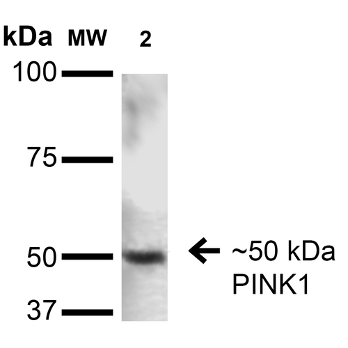 Western Blot analysis of Rat Brain showing detection of ~50 kDa PINK1 protein using Mouse Anti-PINK1 Monoclonal Antibody, Clone S4-15 (56548). Lane 1: Molecular Weight Ladder. Lane 2: Rat Brain. Load: 15 µg. Block: 2% BSA and 2% Skim Milk in 1X TBST. Primary Antibody: Mouse Anti-PINK1 Monoclonal Antibody (56548) at 1:200 for 16 hours at 4°C. Secondary Antibody: Goat Anti-Mouse IgG: HRP at 1:1000 for 1 hour RT. Color Development: ECL solution for 6 min in RT. Predicted/Observed Size: ~50 kDa.
 - -
- -
Antibody DetailsProduct DetailsReactive Species Human ⋅ Mouse ⋅ Rat Host Species Mouse Immunogen Fusion protein corresponding to aa 112-496 (cytoplasmic C-terminus) of human PINK1. This sequence is 82 Product Concentration 1.0 mg/ml Formulation PBS, pH 7.4, 0.1% sodium azide, 50% glycerol. State of Matter Liquid Product Preparation Purified by Protein G affinity chromatography Storage and Handling This product is stable for at least one (1) year at -20°C. Regulatory Status For in vitro investigational use only. Not intended for therapeutic or diagnostic procedures. Country of Origin USA Shipping Next Day 2-8°C Applications and Recommended Usage? Quality Tested by Leinco Immunoblotting: use at 1-5ug/mL. A band of ~50kDa is detected.
Immunofluorescence: use at 10ug/mL. These are recommended concentrations. Enduser should determine optimal concentrations for their application. Each investigator should determine their own optimal working dilution for specific applications. See directions on lot specific datasheets, as information may periodically change. DescriptionDescriptionSpecificity This antibody recognizes human, mouse, and rat PINK1. Background PINK1 (PTEN-induced putative kinase 1) is a mitochondrial serine/threonine kinase that maintains mitochondrial function and integrity, provides protection against mitochondrial dysfunction during cellular stress, and is involved in the clearance of damaged mitochondria via selective autophagy. PINK1 is synthesized as a 63kDa protein which undergoes proteolytic processing to yield two cleaved forms, ~55kDa and ~42kDa. PINK1 may be localized to the outer mitochondrial membrane with its kinase domain facing the cytosol which would explain the observed physical interaction between PINK1 and cytosolic E3 ubiquitin ligase Parkin. NCBI Gene Bank ID UniProt.org Research Area Neuroscience References & CitationsTechnical ProtocolsICC IF   Certificate of Analysis |
Formats Available
 Products are for research use only. Not for use in diagnostic or therapeutic procedures.