

Rat TNF-α ELISA Development Kit


Data
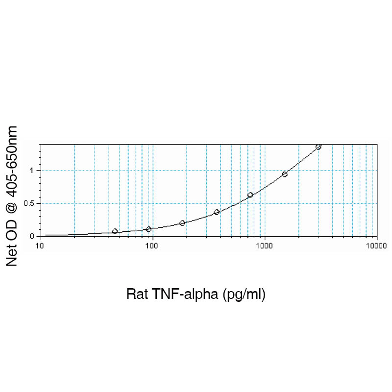
- -
- -
Product DetailsDescription Rat TNF-α ELISA Development Kit contains the key components required for the quantitative measurement of natural and/or recombinant TNF-α in a sandwich ELISA format. Using the ELISA protocol described below, this kit provides sufficient reagents to assay TNF-α in approximately 1500 ELISA plate wells. Materials Provided 1.) Capture Antibody 2.) Detection Antibody 3.) Standard 4.) StreptAvidin-HRP 5.) TMB Liquid Substrate "Ready to Use" Other Materials and Solutions Required Additional Required Materials ELISA microplates (Thermo Fisher Cat. # 456529) BSA (Sigma Cat. # A-7030) Dulbecco’s PBS (DPBS) [10x] (Leinco Product No. D388) Stop Solution: 450 nm Stop Reagent for TMB Microwell (Leinco Product No. T125) Required Solutions PBS: dilute 10xPBS to 1xPBS, pH 7.2, in sterile water Wash Buffer: 0.05% Tween-20 in PBS (Leinco Product No. W101) Reagent Diluent: 1.0% BSA in PBS* Blocking Buffer 1.0% BSA in PBS (Leinco Product No. B395) Note: Other acceptable blocking buffers such as Leinco's Ultra-FISH Block (Leinco Product No. B396) or Ultra-Sythetic Block (Leinco Product No. B397) may be used for assay optimization. Precautions Some of the required components may contain acid and/or cause allergic reactions. Breathing in product mist or fumes should be avoided. Wear protective gloves, clothing, eye, and face protection. Wash hands thoroughly after handling. Please refer to the MSDS on our website prior to use. Capture Antibody Mouse Anti-Rat TNF-α Capture Antibody: Centrifuge vial prior to opening. Reconstitute in 0.5 mL sterile PBS. Refer to the lot-specific datasheet for amount supplied and dilute in PBS without carrier protein to the working concentration indicated on the C of A. Following reconstitution the capture antibody may be stored at 2 – 8°C for up to 6 months. For long term storage, it is recommended to aliquot into working volumes and store at -70°C in a manual defrost freezer. Avoid repeated freeze and thaw cycles. Biotin Detection Antibody Biotinylated Goat Anti-Rat TNF-α Detection Antibody: Refer to the lot-specific datasheet for amount supplied. Centrifuge vial prior to opening. Reconstitute with 1.0 mL of Reagent Diluent. Dilute in Reagent Diluent to the working concentration indicated on the C of A. Detection antibodies may be stored at 2 – 8°C for up to 6 months. For long term storage, it is recommended to aliquot into working volumes and store at -70°C in a manual defrost the freezer. Avoid repeated freeze and thaw cycles. Recombinant Standards E. coli - expressed Recombinant Rat TNF-α Standard: Centrifuge vial prior to opening. Reconstitute each vial with 0.5 mL of Reagent Diluent. Refer to the lot-specific datasheet for amount supplied. The rProtein may be stored at 2 – 8°C for one (1) month or aliquoted and stored at -70°C for up to three months in a manual defrost freezer. Avoid repeated freeze and thaw cycles. Substrate TMB Liquid Substrate "Ready to Use" (TMB Substrate should be at ambient temperature prior to use): 60.0 mL of TMB HRP Microwell Substrate Standard Kinetic One Component "Ready Use" (Leinco Product No. T118) is provided. The high quality of the substrate can be preserved by storing at temperatures between 2 – 8ºC. When properly stored, TMB Microwell Substrate is stable for a minimum of 48 months from the manufactured date. Plate Preparation 1. Dilute the capture antibody to the working concentration in PBS without carrier protein and immediately add 100 μL to each ELISA plate well. Seal the plate and incubate overnight at room temperature. 2. Aspirate the wells to remove liquid and wash the plate 4 times using 300-400 μL of wash buffer per well. Note: We recommend using an autowasher, although a squirt bottle or manifold dispenser would suffice. 3. After the last wash, invert plate to remove residual buffer and blot on paper towel. 4. Add 300 μL block buffer to each well and incubate for at least 1 hour at room temperature. 5. Aspirate and wash plate 4 times. NOTE: Complete removal of liquid at each step is essential for good performance and sensitivity of assay. Assay Procedure Standard/Sample: Add 100 μL of the working dilution with reagent dilution standard or sample to each well (duplicate recommended). Cover plate with an adhesive plate cover and incubate at room temperature for at least 2 hours. Detection: Aspirate and wash plate 4 times. Add 100 μL of the detection antibody, diluted in Reagent Diluent to each well. Cover with a new adhesive plate cover and incubate at room temperature for 2 hours. StreptAvidin-HRP Conjugate: Aspirate and wash plate 4 times. Add 100 μL of the working dilution (the dilution factor may require optimization) to each well. Cover and incubate at room temperature for 20-30 minutes. Exposure to direct light should be avoided. TMB Liquid Substrate: Aspirate and wash plate 4 times. Add 100 μL of TMB HRP Microwell Substrate Standard Kinetic One Component "Ready Use" (Leinco Product No. T118) to each well. Incubate at room temperature for 20-30 minutes and monitor color development. Exposure to direct light should be avoided. Stop Solution: Add 50-100 μL of Stop Solution (Leinco Product No. T125) to each well. Monitor color development with an ELISA plate reader at 450 nm with wavelength correction set at 540 nm or 570 nm. BackgroundCD120 can refer to two members of the tumor necrosis factor receptor superfamily- CD120a (TNFR1) or CD120b (TNFR2). CD120a is a 55kD Type I transmembrane protein receptor that binds both TNF-α and TNF-β (LT-α). In association with TRADD and RIP, the receptor crosslinking induced by TNF-α or TNF-β trimers is vital for signal transduction, leading to apoptosis, NF-B activation, increased expression of proinflammatory genes, tumor necrosis, and cell differentiation depending on cell type and differentiation state. CD120b is a 75 kD type I transmembrane protein that binds both TNF-α and TNF-β. In conjunction with TRAF1 and TRAF2, the receptor crosslinking induced by TNF-α or TNF-β trimers is critical for signal transduction that may lead to apoptosis, NF-kB activation, increased expression of proinflammatory genes, tumor necrosis, and cell differentiation depending on cell type and differentiation state. TNF-α is a 17.5 kD protein that mediates inflammation and immunity caused by the invasion of viruses, bacteria, and parasites by initiating a cascade of cytokines that increase vascular permeability, thus bringing macrophages and neutrophils to the site of infection. TNF-α secreted by the macrophage causes the blood to clot which provides containment of the infection. TNF-α binding to surface receptors brings about various biologic activities that include cytolysis and cytostasis of many tumor cell lines In vitro, hemorraghic necrosis of tumors In vivo, increased fibroblast proliferation, and enhanced chemotaxis and phagocytosis in neutrophils. TNF-β (LT-α) is a 25 kD protein that has a significant impact on the maintenance of the immune system including the development of secondary lymphoid organs. TNF-β has dual functions. It may function to prevent growth of cancer cells or it may facilitate the development of tumors. TNF-β is involved in the regulation of cell survival, proliferation, differentiation, and apoptosis and, if unregulated, can result in a constantly active signaling pathway, resulting in uncontrolled cellular growth and creation of tumors. Additionally, TNF-β is involved in innate immune regulation and has been shown to prevent tumor growth and obliterate cancerous cell lines. References & Citations1. El-Harith el-HA et al. (2004) Saudi Med J. 25: 135
2. Adolf GR et al. (1990) Infec Immun. 58: 3996 |
Related Products
- -
- -
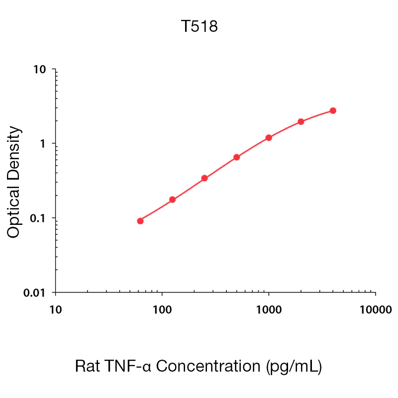
Prod No. | Description |
|---|---|
T518 | |
T519 |
 Products are for research use only. Not for use in diagnostic or therapeutic procedures.