

Recombinant Human FGF-8a


Data
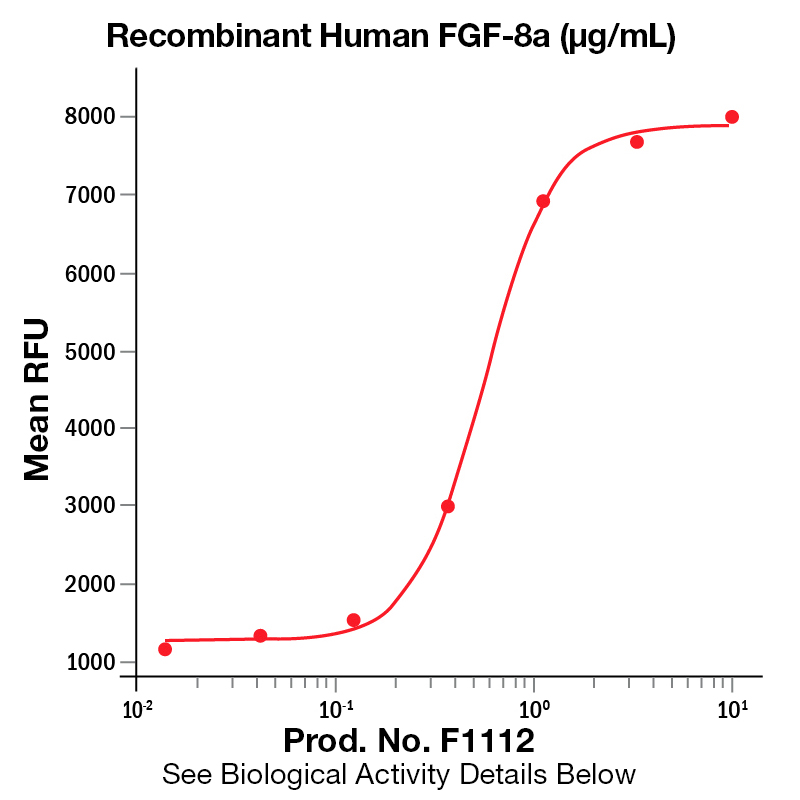
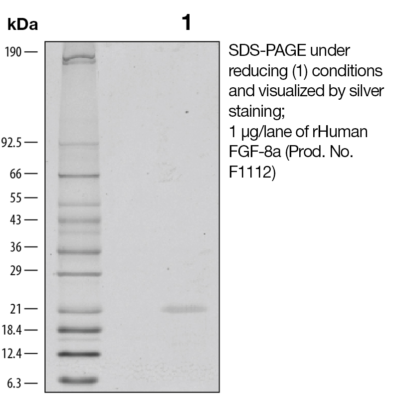
BackgroundFibroblast growth factor-8 (FGF-8), also known as AIGF and HBGF, is a heparin binding growth factor belonging to the FGF family (1). Proteins of this family play a central role during prenatal development and postnatal growth and regeneration of a variety of tissues, by promoting cellular proliferation and differentiation (2). Alternate splicing of FGF-8 mRNA creates eight secreted isoforms (a-h) in mice and four (a, b, e and f) in humans (3). FGF-8a expands the midbrain in transgenic mice, while FGF-8b transforms the midbrain into cerebellum. FGF-8 activates the “c” splice forms of receptors FGF R2, FGF R3 and FGF R4, with differential activity among the FGF-8 isoforms. Overexpression of FGF-8 has been shown to increase tumor growth and angiogenesis. FGF-8b shows the strongest receptor affinity and oncogenic transforming capacity, although isoforms a and e have been found in human tumors (4). The adult expression of FGF-8 is restricted to testes and ovaries. Protein DetailsPurity >95% by SDS-PAGE and analyzed by silver stain. Endotoxin Level <0.01 EU/µg as determined by the LAL method Biological Activity The biological activity of Human FGF-8a was determined by a cell proliferation assay using NR6R-3T3 mouse fibroblasts. The expected ED<sub>50</sub>=0.4 - 2µg/ml in the presence of 10µg/ml heparin. Protein Accession No. Amino Acid Sequence qhvreqslvt dqlsrrlirt yqlysrtsgk hvqvlankri namaedgdpf aklivetdtf gsrvrvrgae tglyicmnkk gkliaksngk gkdcvfteiv lennytalqn akyegwymaf trkgrprkgs ktrqhqrevh fmkrlprghh tteqslrfef lnyppftrsl rgsqrtwape pr N-terminal Sequence Analysis Met State of Matter Lyophilized Predicted Molecular Mass The predicted molecular weight of Recombinant Human FGF-8a is Mr 21 kDa. Predicted Molecular Mass 21 Formulation This recombinant protein was lyophilized from a 0.2 μm filtered solution in MOPS, EDTA, Dithiothreitol (DTT), and sodium sulphate (Na2SO4). Storage and Stability This lyophilized protein is stable for six to twelve months when stored desiccated at -20°C to -70°C. After aseptic reconstitution, this protein may be stored at 2°C to 8°C for one month or at -20°C to -70°C in a manual defrost freezer. Avoid Repeated Freeze Thaw Cycles. See Product Insert for exact lot specific storage instructions. Country of Origin USA Shipping Next Day Ambient NCBI Gene Bank Applications and Recommended Usage ? (Quality Tested by Leinco) ELISA Sandwich: This antibody is useful as the capture antibody in a sandwich ELISA. The suggested coating concentration is 5 µg/ml (100 µl/well) µg/ml. Flow Cytometry: PN:A106 Flow Cytometry: It is recommended to use the indirect method for signal enhancement when enumerating cells expressing CXCR5. A suggested method would be to stain cells expressing CXCR5 with approximately 10 µl per test. A typical test sample constitutes approximately 50 µl of packed whole blood or 1 x 10⁵ continuous passage or activated cell cultures that have been centrifuged at 500 X g for five minutes. Labeling of the cells with the biotin conjugate should be followed by PN:A104, resuspended in 200-400 µl of 1X PBS. Leinco Protein AdvisorPowered by AI: AI is experimental and still learning how to provide the best assistance. It may occasionally generate incorrect or incomplete responses. Please do not rely solely on its recommendations when making purchasing decisions or designing experiments. Recombinant Human FGF-8a is a valuable tool for research due to its well-documented roles in regulating key cellular processes and developmental pathways. Here are the main reasons to use Recombinant Human FGF-8a in your research applications: 1. Regulation of Embryogenesis and DevelopmentFGF-8a is essential for embryonic development, including the formation of the midbrain/hindbrain, limbs, eyes, ears, and heart. It mediates epithelial-mesenchymal transitions and plays an organizing and inducing role during gastrulation. This makes it critical for studies on developmental biology and organogenesis. 2. Cell Proliferation, Differentiation, and MigrationFGF-8a is a potent mitogen that stimulates cell proliferation, differentiation, and migration. It activates Ras/MAPK signaling pathways, which are central to these processes. This property is useful for experiments involving stem cell expansion, tissue regeneration, and cancer research. 3. Neural Stem Cell and iPSC DifferentiationFGF-8a is widely used to differentiate induced pluripotent stem cells (iPSCs), embryonic stem cells (ESCs), and neural stem cells. It supports the generation of specific neural lineages and is frequently applied in protocols for modeling neurological diseases and developmental disorders. 4. High Purity and Biological ActivityRecombinant Human FGF-8a is typically available with high purity (>95–98%) and low endotoxin levels, ensuring reliable and reproducible results in sensitive cell-based assays. Its biological activity is validated using standardized proliferation assays (e.g., NR6R-3T3 or BALB/c 3T3 cells), with ED₅₀ values in the range of 0.1–2 μg/mL. 5. Relevance to Disease ModelsFGF-8a has been implicated in tumor growth, angiogenesis, and certain cancers (e.g., prostate, breast, ovarian). Its expression is also linked to tissue repair and wound healing, making it relevant for studies on cancer biology, regenerative medicine, and tissue engineering. 6. Well-Characterized and Widely CitedRecombinant Human FGF-8a is supported by extensive scientific literature and product validation data. Its use is documented in numerous peer-reviewed publications, ensuring that your research is built on a solid and credible foundation. 7. Versatile ApplicationsIt is suitable for a wide range of applications, including:
8. Species CompatibilityRecombinant Human FGF-8a is active in human, mouse, and other mammalian cell systems, making it broadly applicable across different experimental models. In summary, Recombinant Human FGF-8a is a versatile, well-characterized growth factor that supports a wide array of research applications, particularly in developmental biology, stem cell research, and disease modeling. Its high purity, biological activity, and relevance to key cellular processes make it an essential reagent for advanced scientific studies. Recombinant Human FGF-8a can be used as a standard for quantification or calibration in ELISA assays, provided it is of high purity, well-characterized, and compatible with your assay system. Key considerations and supporting details:
Summary Table: Key Requirements for Using Recombinant FGF-8a as an ELISA Standard
Conclusion: Recombinant Human FGF-8a has been validated for several key applications in published research, primarily in the context of cell culture, stem cell biology, and developmental studies. The main applications supported by published data include:
These applications are supported by primary research articles and product validation data from multiple sources, confirming the utility of Recombinant Human FGF-8a in a wide range of scientific studies. To reconstitute and prepare Recombinant Human FGF-8a protein for cell culture experiments, dissolve the lyophilized protein in sterile water or buffer to a concentration of at least 100 μg/mL, incubate at room temperature for 20 minutes, and avoid vortexing to preserve biological activity. After reconstitution, further dilute in cell culture medium or buffer containing a carrier protein such as 0.1% BSA or 10% FBS to stabilize the protein for experimental use. Step-by-step protocol:
Summary of key points:
These steps ensure optimal solubility, stability, and biological activity of recombinant FGF-8a for cell culture experiments. References & Citations1. Gemel, J. et al. (1996) Genomics 35:253 2. Ruess, B. et al. (2003) Cell Tissue Res. 313:139 3. Tanaka, S. et al. (2001) Digest. Dis. Sci. 46:1016 4. Olsen, SK. et al. (2006) Genes Dev. 20:185 Technical Protocols  Certificate of AnalysisIMPORTANT Use lot specific datasheet for all technical information pertaining to this recombinant protein. |
Related Products
 Products are for research use only. Not for use in diagnostic or therapeutic procedures.