HEK293-derived human Galectin-2 protein Met1-Glu132 (Accession # P05162)
N-terminal Sequence Analysis
Met1 & Thr2
State of Matter
Lyophilized
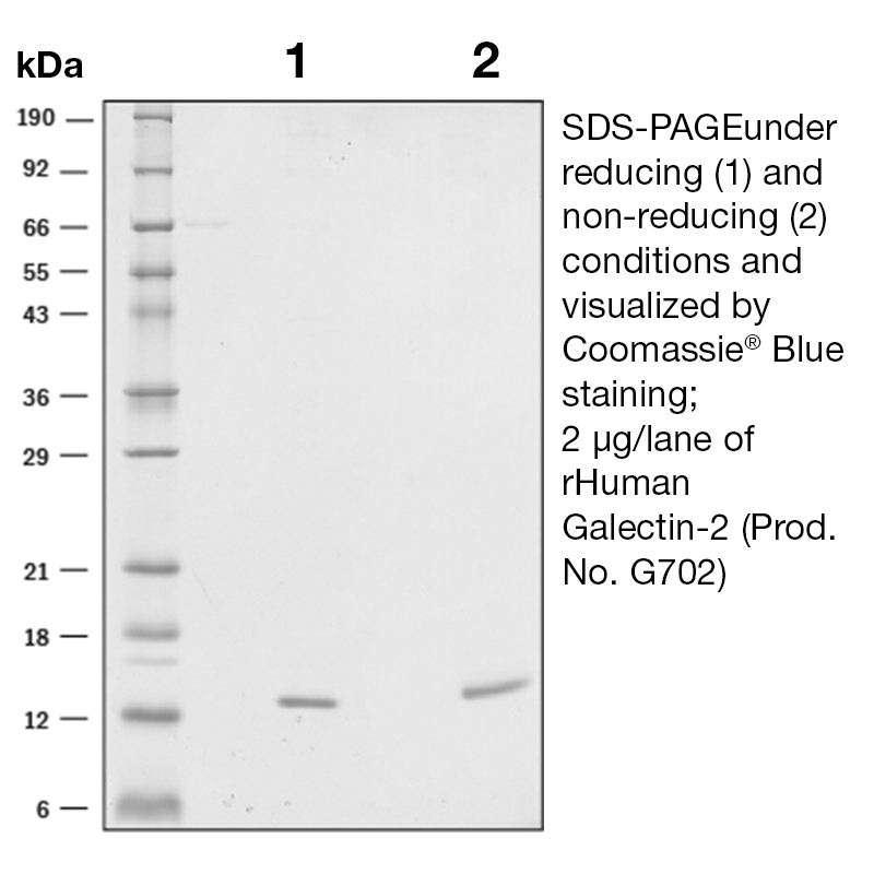
Predicted Molecular Mass
The predicted molecular weight of Recombinant Human Galectin-2 is Mr 14 kDa.
Predicted Molecular Mass
14
Formulation
This recombinant protein was 0.2 µm filtered and lyophilized from 0.2 μm filtered solution in HEPES, NaCl, TCEP, PEG and Trehalose.
Storage and Stability
This lyophilized protein is stable for six to twelve months when stored desiccated at -20°C to -70°C. After aseptic reconstitution, this protein may be stored at 2°C to 8°C for one month or at -20°C to -70°C in a manual defrost freezer. Avoid Repeated Freeze Thaw Cycles. See Product Insert for exact lot specific storage instructions.
Applications and Recommended Usage ? (Quality Tested by Leinco)
ELISA Sandwich: This antibody is useful as the capture antibody in a sandwich ELISA. The suggested coating concentration is 5 µg/ml (100 µl/well) µg/ml. Flow Cytometry: PN:A106 Flow Cytometry: It is recommended to use the indirect method for signal enhancement when enumerating cells expressing CXCR5. A suggested method would be to stain cells expressing CXCR5 with approximately 10 µl per test. A typical test sample constitutes approximately 50 µl of packed whole blood or 1 x 10⁵ continuous passage or activated cell cultures that have been centrifuged at 500 X g for five minutes. Labeling of the cells with the biotin conjugate should be followed by PN:A104, resuspended in 200-400 µl of 1X PBS.
Leinco Protein Advisor
Powered by AI: AI is experimental and still learning how to provide the best assistance. It may occasionally generate incorrect or incomplete responses. Please do not rely solely on its recommendations when making purchasing decisions or designing experiments.
Recombinant Human Galectin-2 is a versatile tool for research due to its roles in immunology, inflammation, epithelial biology, and disease modeling. Its use enables controlled, reproducible studies of galectin-2’s molecular and cellular functions, which are relevant to several physiological and pathological processes.
Key reasons to use recombinant human galectin-2 in research applications:
Immunomodulation and Inflammation Studies Galectin-2 modulates immune cell function, including monocytes and macrophages, by inducing proinflammatory cytokines (e.g., TNF-α, IL-6, IL-12p40, IFN-β) and influencing anti-arteriogenic pathways via the CD14/TLR4 axis. It also affects T cell cytokine secretion and promotes immune cell apoptosis, making it valuable for dissecting immune response mechanisms.
Epithelial Barrier and Wound Healing Research Galectin-2 enhances epithelial integrity by interacting with β-catenin and E-cadherin, promoting cell adhesion and migration. It has been shown to preserve mucosal barrier function and support wound healing in gastrointestinal models.
Disease Modeling Altered galectin-2 expression is implicated in inflammatory bowel disease, cardiovascular disease, cancer, and pregnancy disorders. Recombinant protein allows for mechanistic studies and therapeutic target validation in these contexts.
Apoptosis and Cell Death Pathways Galectin-2 induces apoptosis in T cells, neutrophils, and monocytes, providing a model to study cell death pathways relevant to immune regulation and disease pathogenesis.
Cancer and Tumor Microenvironment Galectin-2 participates in tumor progression, metastasis, and the regulation of inflammation within the tumor microenvironment, making it useful for cancer biology research.
Standardization and Reproducibility Recombinant production ensures batch-to-batch consistency, purity, and the absence of contaminating factors, which is critical for reproducible experimental results.
Functional Assays Recombinant galectin-2 can be used in cell-based assays (e.g., cytokine release, apoptosis, migration), biochemical binding studies, and as a control or stimulus in immunological experiments.
In summary, recombinant human galectin-2 is a powerful reagent for dissecting its diverse biological roles, enabling precise, hypothesis-driven research in immunology, epithelial biology, and disease modeling.
Yes, recombinant human Galectin-2 (GAL2) can be used as a standard for quantification or calibration in ELISA assays, provided that the ELISA kit is designed to detect both recombinant and native forms of Galectin-2.
Key Points to Consider:
Compatibility with ELISA Kit: Most commercial ELISA kits for human Galectin-2 are validated for use with recombinant standards. For example:
Rockland’s Human Galectin-2 ELISA Kit (KOA0754) explicitly states that it detects both natural and recombinant human Galectin-2.
Cohesion Biosciences and ELK Biotech kits also mention detection of recombinant Galectin-2.
However, some kits (e.g., MyBioSource and Cloud-Clone) specify that they are designed for native Galectin-2 and may not be optimal for recombinant protein detection.
Standard Preparation:
Use a purified recombinant Galectin-2 protein (preferably with a known concentration and purity).
Prepare serial dilutions of the recombinant standard to generate a standard curve within the detection range of your ELISA kit.
Ensure the recombinant protein is compatible with the assay buffer and does not contain interfering substances (e.g., BSA or other carriers, unless specified by the kit manufacturer).
Validation:
Always validate the use of your recombinant standard by checking linearity and recovery in your specific assay.
If possible, compare results with a native standard or a kit-provided standard to confirm accuracy.
Recommendations:
Check the ELISA kit manual for specific guidance on standard selection.
Use recombinant Galectin-2 expressed in a compatible system (e.g., E. coli or HEK-293, depending on kit requirements).
Avoid kits that explicitly state they are not suitable for recombinant protein detection unless you have validated the use of recombinant standards.
In summary, recombinant human Galectin-2 is generally suitable as an ELISA standard, but always confirm compatibility with your specific kit and validate performance in your assay.
Recombinant Human Galectin-2 has been validated for several key applications in published research, primarily in studies of immunology, cell biology, and disease models.
Validated Applications:
Immunoblotting and Serodiagnosis: Recombinant galectin-2 has been used as an antigen in immunoblot assays to detect specific antibodies in human serum, particularly for the diagnosis of parasitic infections such as angiostrongyliasis. This application demonstrates high sensitivity and specificity for distinguishing disease states.
Cell Adhesion and Migration Assays: Galectin-2 has been shown to enhance adhesion and migration of human colon cancer cells (Caco-2) and non-transformed rat intestinal epithelial cells (IEC-6) by interacting with β-catenin and E-cadherin complexes. These assays are used to study cell-cell interactions and wound healing mechanisms.
In Vivo Disease Models: Administration of recombinant galectin-2 in mouse models has been validated for studying gastrointestinal wound healing, colitis, and epithelial barrier integrity. Exogenous galectin-2 preserves crypt architecture, modulates cytokine release (increasing IL-10, decreasing IL-6 and IL-12p70), and induces T cell apoptosis in the lamina propria.
Macrophage Polarization and Arteriogenesis: Recombinant galectin-2 has been used in vitro and in vivo to study its effects on monocyte/macrophage phenotype. It induces a proinflammatory, anti-arteriogenic phenotype via CD14/TLR4 signaling, and its inhibition promotes a proarteriogenic M2 phenotype, improving collateral vessel growth in murine models of arterial stenosis.
Apoptosis Induction: Recombinant galectin-2 has been validated for inducing apoptosis in various cell types, including T cells and neutrophils, both in vitro and in vivo.
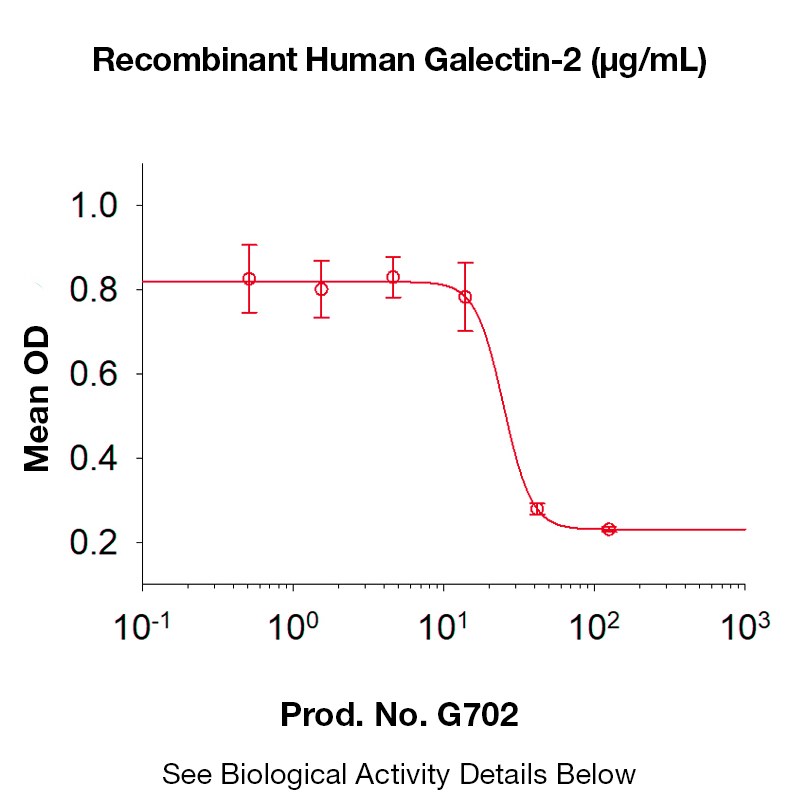
Agglutination Assays: Recombinant human galectin-2 has been shown to agglutinate human red blood cells, with a defined ED50, supporting its use in lectin activity assays.
Additional Context:
Cancer Research: Galectin-2 is implicated in tumor progression, immune response modulation, and pre-mRNA splicing, and recombinant protein is used to study these processes.
Protein Interaction Studies: Recombinant galectin-2 is used in binding assays to characterize interactions with carbohydrates and peptides, relevant for structural biology and ligand specificity studies.
Inflammatory Disease Models: Altered galectin-2 expression and recombinant protein administration have been studied in models of inflammatory bowel disease, coronary artery disease, rheumatoid arthritis, and pregnancy disorders.
Summary Table of Validated Applications
Application Type
Experimental Contexts/Models
Key Outcomes/Readouts
Immunoblotting/Serodiagnosis
Human serum, parasitic infection diagnosis
Sensitivity, specificity, accuracy
Cell Adhesion/Migration Assays
Cancer/epithelial cell lines
Adhesion, migration, wound healing
In Vivo Disease Models
Mouse colitis, epithelial integrity
Cytokine modulation, apoptosis
Macrophage Polarization
Murine arteriogenesis, monocyte assays
M1/M2 phenotype, vessel growth
Apoptosis Induction
T cells, neutrophils, in vitro/in vivo
Cell death quantification
Agglutination Assays
Human red blood cells
Lectin activity, ED50 determination
Protein Interaction Studies
Carbohydrate/peptide binding assays
Affinity, specificity
These applications are supported by peer-reviewed studies and demonstrate the utility of recombinant human galectin-2 in diverse experimental protocols relevant to immunology, cell biology, and disease research.
To reconstitute and prepare Recombinant Human Galectin-2 protein for cell culture experiments, dissolve the lyophilized protein in sterile water or buffer to a concentration of at least 100 μg/mL, incubate for 20 minutes to ensure complete dissolution, and then dilute to your working concentration using sterile cell culture-compatible buffer (such as PBS with 0.1% BSA) under aseptic conditions.
Detailed protocol and best practices:
Reconstitution:
Use sterile, endotoxin-free water or sterile PBS as the initial solvent.
Add enough solvent to achieve a concentration of at least 100 μg/mL. Some protocols recommend up to 200 μg/mL.
Gently mix (do not vortex vigorously) and incubate at room temperature for at least 20 minutes to ensure the protein is fully dissolved.
If the protein is difficult to dissolve, gentle pipetting or brief low-speed vortexing may help, but avoid foaming.
Buffer considerations:
For cell culture, it is best to use sterile PBS or cell culture medium as the final buffer. If the protein was reconstituted in water, dilute it into PBS or medium for use.
To enhance stability and prevent adsorption, add 0.1% BSA (bovine serum albumin) to the buffer if compatible with your assay.
Avoid high concentrations of reducing agents or detergents unless required for your specific application.
Sterility and endotoxin:
Ensure all solutions and containers are sterile.
If the protein will be used in sensitive cell assays, confirm that it is low in endotoxin (LPS). Some protocols recommend passing the protein through an endotoxin removal column if necessary.
Aliquoting and storage:
After reconstitution, aliquot the protein to avoid repeated freeze-thaw cycles.
Store aliquots at -20°C or -80°C for long-term storage; use within one month after reconstitution.
For short-term use (up to 1 week), storage at 2–8°C is acceptable under sterile conditions.
Working solution preparation:
Thaw an aliquot on ice before use.
Dilute to the desired working concentration in cell culture medium immediately before adding to cells.
Typical working concentrations for functional assays range from 0.1–10 μg/mL, but optimize based on your experimental design.
Summary Table:
Step
Recommendation
Solvent
Sterile water or PBS (endotoxin-free)
Initial concentration
≥100 μg/mL (up to 200 μg/mL)
Incubation
20 min at room temperature
Buffer for use
PBS or cell culture medium, optionally with 0.1% BSA
Sterility
Use sterile technique; confirm low endotoxin if needed
Storage
Aliquot and store at -20°C or -80°C; avoid repeated freeze-thaw
Working dilution
Prepare fresh in cell culture medium; typical range 0.1–10 μg/mL
References:
Reconstitution and storage recommendations.
Endotoxin removal and sterility for cell culture.
General recombinant protein handling for cell culture.
If your protein preparation includes specific additives (e.g., glycerol, DTT), check compatibility with your cell system and consider buffer exchange if necessary. Always consult the product datasheet for any protein-specific instructions.


 Products are for research use only. Not for use in diagnostic or therapeutic procedures.