GDF-11/BMP-11 protein Asn299-Ser407, with an N-terminal Met (Accession #: O95390)
State of Matter
Lyophilized
Predicted Molecular Mass
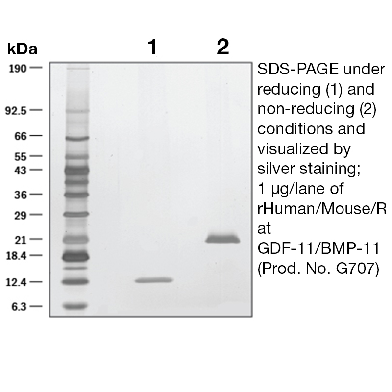
The predicted molecular weight of Recombinant Human GDF11, BMP11 is Mr 12.6 kDa.
Predicted Molecular Mass
12.6
Formulation
This BMP-11 proteins was lyophilized from a 0.2 μm filtered solution in Acetonitrile and TFA without BSA as a carrier protein.
Storage and Stability
This lyophilized protein is stable for six to twelve months when stored desiccated at -20°C to -70°C. After aseptic reconstitution, this protein may be stored at 2°C to 8°C for one month or at -20°C to -70°C in a manual defrost freezer. Avoid Repeated Freeze Thaw Cycles. See Product Insert for exact lot specific storage instructions.
Applications and Recommended Usage ? (Quality Tested by Leinco)
ELISA Sandwich: This antibody is useful as the capture antibody in a sandwich ELISA. The suggested coating concentration is 5 µg/ml (100 µl/well) µg/ml. Flow Cytometry: PN:A106 Flow Cytometry: It is recommended to use the indirect method for signal enhancement when enumerating cells expressing CXCR5. A suggested method would be to stain cells expressing CXCR5 with approximately 10 µl per test. A typical test sample constitutes approximately 50 µl of packed whole blood or 1 x 10⁵ continuous passage or activated cell cultures that have been centrifuged at 500 X g for five minutes. Labeling of the cells with the biotin conjugate should be followed by PN:A104, resuspended in 200-400 µl of 1X PBS.
Leinco Protein Advisor
Powered by AI: AI is experimental and still learning how to provide the best assistance. It may occasionally generate incorrect or incomplete responses. Please do not rely solely on its recommendations when making purchasing decisions or designing experiments.
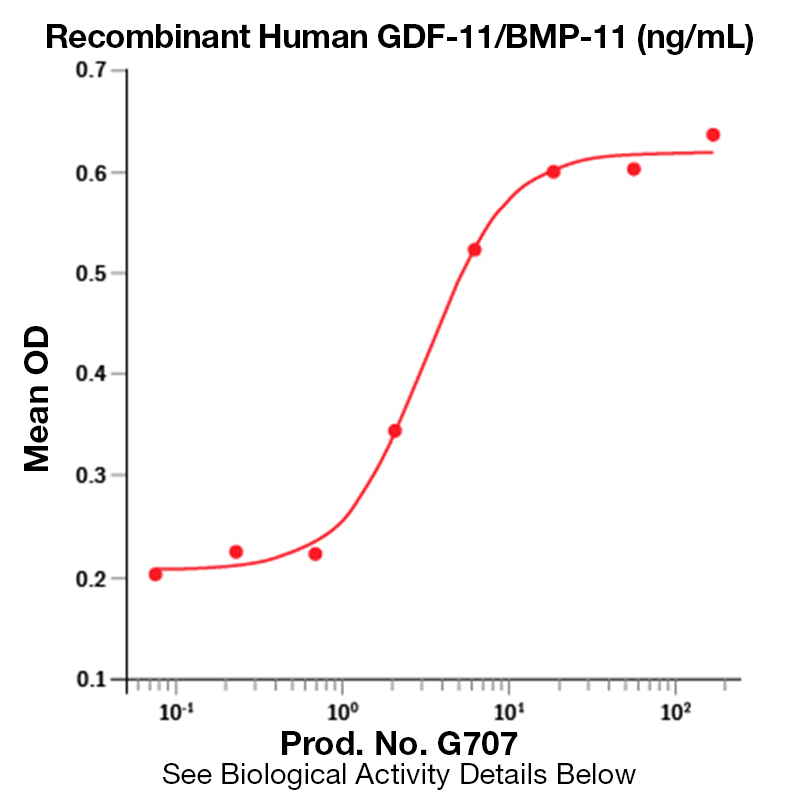
Using Recombinant Human GDF-11 in research applications is valuable due to its demonstrated roles in tissue regeneration, anti-aging effects, neurogenesis, vascular remodeling, and modulation of inflammation across multiple preclinical models.
Key scientific rationales for using recombinant GDF-11 include:
Regeneration and Anti-Aging: GDF-11 levels decline with age, and supplementation with recombinant GDF-11 has been shown to reverse age-related cardiac hypertrophy, restore genomic integrity in muscle stem cells, and improve muscle regeneration, exercise endurance, and neuromuscular function in aged animal models.
Neuroprotection and Neurogenesis: Recombinant GDF-11 promotes neurogenesis, enhances neurovascular health, and improves cognitive performance in models of neurodegenerative diseases and stroke. It increases the number of neural stem cells and brain endothelial cells, and stimulates angiogenesis, which is critical for brain repair after injury.
Vascular and Endothelial Effects: GDF-11 improves endothelial cell function, promotes neovascularization, and accelerates wound healing by enriching endothelial progenitor cells. It also reduces atherosclerotic plaque formation and mitigates endothelial dysfunction in diabetic and ischemic models.
Immunomodulation: GDF-11 can shift macrophage polarization from a pro-inflammatory (M1) to an anti-inflammatory (M2) phenotype, thereby reducing inflammation and supporting tissue repair.
Disease Models: Recombinant GDF-11 has shown therapeutic potential in preclinical models of cardiac fibrosis, ischemic stroke, Alzheimer’s disease, diabetes-related vascular complications, and several cancers, making it a versatile tool for studying disease mechanisms and potential interventions.
Experimental applications include:
In vitro assays for cell differentiation, proliferation, and migration.
In vivo studies for tissue regeneration, neuroprotection, and metabolic regulation.
Mechanistic studies of TGF-β signaling pathways, especially SMAD2/3 activation.
Considerations: Batch-to-batch variability and dosing regimens can affect experimental outcomes, so rigorous controls and validation are recommended. Some controversies remain regarding the extent and context of GDF-11’s effects, but its broad biological activity and reproducible benefits in multiple systems make it a compelling reagent for research into regeneration, aging, and disease.
In summary, recombinant human GDF-11 is a powerful tool for investigating mechanisms of tissue repair, aging, neurovascular health, and immune modulation, with broad translational relevance across regenerative medicine and disease modeling.
Yes, recombinant human GDF-11 can be used as a standard for quantification or calibration in ELISA assays, provided it is compatible with the specific ELISA kit and protocol. Many commercial ELISA kits for GDF-11 are validated to detect both endogenous (natural) and recombinant forms of human GDF-11, and their protocols routinely use recombinant GDF-11 as the standard for generating calibration curves.
Key considerations for use:
Standard Curve Preparation: ELISA kits typically include recombinant human GDF-11 as the standard, and the standard curve is generated by serial dilution of this protein within the assay’s detection range (e.g., 31.2 pg/mL to 2000 pg/mL).
Specificity: Most validated kits report high specificity for human GDF-11, with minimal cross-reactivity to related proteins, ensuring accurate quantification of recombinant GDF-11.
Bioactivity and Purity: Recombinant GDF-11 used as a standard should be of high purity and bioactivity, as confirmed by SDS-PAGE and functional assays.
Kit Compatibility: Always confirm that your ELISA kit is validated for recombinant GDF-11. Some kits may specify that they are not recommended for recombinant proteins, but most GDF-11 ELISA kits are designed to quantify both natural and recombinant forms.
Best Practices:
Use the recombinant GDF-11 standard provided or recommended by the ELISA kit manufacturer to ensure compatibility with the assay’s antibodies and detection system.
Prepare the standard curve fresh for each assay, following the dilution instructions in the kit manual.
Validate the standard curve in your specific matrix (e.g., serum, plasma, cell culture media) to account for potential matrix effects.
Confirm the concentration and integrity of your recombinant GDF-11 standard by independent quantification (e.g., BCA assay, SDS-PAGE) if using a source other than the kit-provided standard.
Limitations:
Some ELISA kits may have limited cross-reactivity testing, so if your recombinant GDF-11 differs in sequence or post-translational modifications from the endogenous protein, verify its recognition by the kit antibodies.
Always consult the specific ELISA kit manual for recommendations regarding recombinant protein standards, as a minority of kits may advise against their use for calibration.
Summary Table: Use of Recombinant Human GDF-11 as ELISA Standard
Kit Type/Protocol
Recombinant GDF-11 as Standard
Notes/Limitations
Most commercial GDF-11 ELISA kits
Yes
Standard curve routinely prepared with recombinant protein
Kits with limited cross-reactivity validation
Caution
Confirm recognition of your recombinant protein
Custom ELISA development
Yes, if antibodies recognize recombinant GDF-11
Validate standard curve and specificity
In conclusion, recombinant human GDF-11 is widely accepted and scientifically appropriate as a standard for ELISA quantification, provided it matches the kit’s requirements and is properly validated.
Recombinant Human GDF-11 has been validated for a range of applications in published research, primarily in functional and blocking assays, as well as in diverse in vitro and in vivo models investigating its biological effects.
Key validated applications include:
Functional Assays: Recombinant GDF-11 is widely used to assess its biological activity on various cell types, including neural stem cells, endothelial cells, and fibroblasts, to study effects such as neurogenesis, angiogenesis, and extracellular matrix production.
Blocking Assays: It is used to block or neutralize endogenous GDF-11 activity in cell-based systems to dissect its specific signaling pathways and physiological roles.
In Vivo Animal Models: Systemic administration of recombinant GDF-11 has been validated in rodent models to study:
Neuroregeneration and Stroke Recovery: Enhances neovascularization, reduces inflammation, promotes neurogenesis, and improves sensorimotor function after ischemic stroke in rats.
Aging and CNS Plasticity: Increases hippocampal neurogenesis, improves vasculature, and enhances neuronal activity and plasticity in aged mice.
Tissue Fibrosis: Stimulates extracellular matrix protein production in fibroblasts, relevant to studies of fibrosis in heart, kidney, and liver.
Endothelial Function and Atherosclerosis: Protects against endothelial injury, reduces atherosclerotic lesion formation, and improves endothelial cell function in models of cardiovascular and metabolic disease.
Stem Cell Regulation: Modulates the function and therapeutic efficacy of mesenchymal and neural stem cells in regenerative medicine contexts.
In Vitro Cell Culture Studies: Used to investigate:
Macrophage Polarization: Facilitates conversion of pro-inflammatory (M1) to anti-inflammatory (M2) macrophages, impacting immune responses.
Metabolic Regulation: Influences glucose metabolism and cholesterol handling in various cell types.
Developmental Biology: Regulates development of olfactory system, retina, pancreas, and axial skeleton patterning.
Summary Table: Validated Applications of Recombinant Human GDF-11
Recombinant GDF-11 is typically expressed in E. coli and validated for activity in both human and rodent systems.
Its effects are often mediated through SMAD2/3 signaling and can be context-dependent, varying by cell type, age, and disease state.
Published studies use a variety of readouts, including immunofluorescence, ELISA, proteomics, and behavioral assays in animals.
If you require protocols or more specific details on a particular application, please specify the context or experimental system of interest.
To reconstitute and prepare Recombinant Human GDF-11 protein for cell culture experiments, dissolve the lyophilized protein in sterile 4 mM HCl or sterile deionized water, depending on the specific formulation and manufacturer’s recommendations. The most common and widely supported protocol is as follows:
Reconstitution concentration: 100–200 μg/mL in sterile 4 mM HCl. Alternatively, some protocols recommend 0.1–1.0 mg/mL in sterile deionized water.
Procedure:
Briefly centrifuge the vial to collect the powder at the bottom before opening.
Add the appropriate volume of sterile 4 mM HCl (or sterile water, if specified) to achieve the desired concentration.
Gently pipette the solution down the sides of the vial to dissolve the protein. Do not vortex.
Allow the vial to sit at room temperature for several minutes to ensure complete dissolution.
Aliquoting and storage: After reconstitution, aliquot the solution to avoid repeated freeze-thaw cycles and store at –20 °C or below. Avoid multiple freeze-thaw cycles to preserve activity.
Dilution for cell culture: Further dilute the reconstituted stock into your cell culture medium immediately before use. If using acidic reconstitution (HCl), ensure the final HCl concentration in the culture medium is negligible and does not affect cell viability. You may dilute the stock into a neutral buffer (e.g., PBS with 0.1% BSA) before adding to cells.
Key technical notes:
Choice of solvent: Most protocols recommend 4 mM HCl for initial reconstitution, especially for carrier-free or highly purified GDF-11, as this helps maintain solubility and stability. Some preparations with carrier protein (e.g., BSA) may allow reconstitution in sterile water.
Concentration: Prepare a concentrated stock (e.g., 100–200 μg/mL or 0.1–1.0 mg/mL) and dilute as needed for your assay.
Handling: Avoid vigorous mixing or vortexing, which can denature the protein.
Sterility: Always use sterile reagents and equipment to prevent contamination in cell culture.
Summary Table:
Step
Recommended Protocol
Solvent
Sterile 4 mM HCl (most common) or sterile water
Stock concentration
100–200 μg/mL (HCl) or 0.1–1.0 mg/mL (water)
Mixing
Gentle pipetting, no vortexing
Storage
Aliquot, –20 °C or below, avoid freeze-thaw cycles
Working dilution
Dilute into culture medium or neutral buffer before use
Always consult the specific product datasheet for your recombinant GDF-11, as formulation and recommended protocols may vary depending on the expression system and presence of carrier proteins.


 Products are for research use only. Not for use in diagnostic or therapeutic procedures.