

Anti-Choline Acetyltransferase


Data
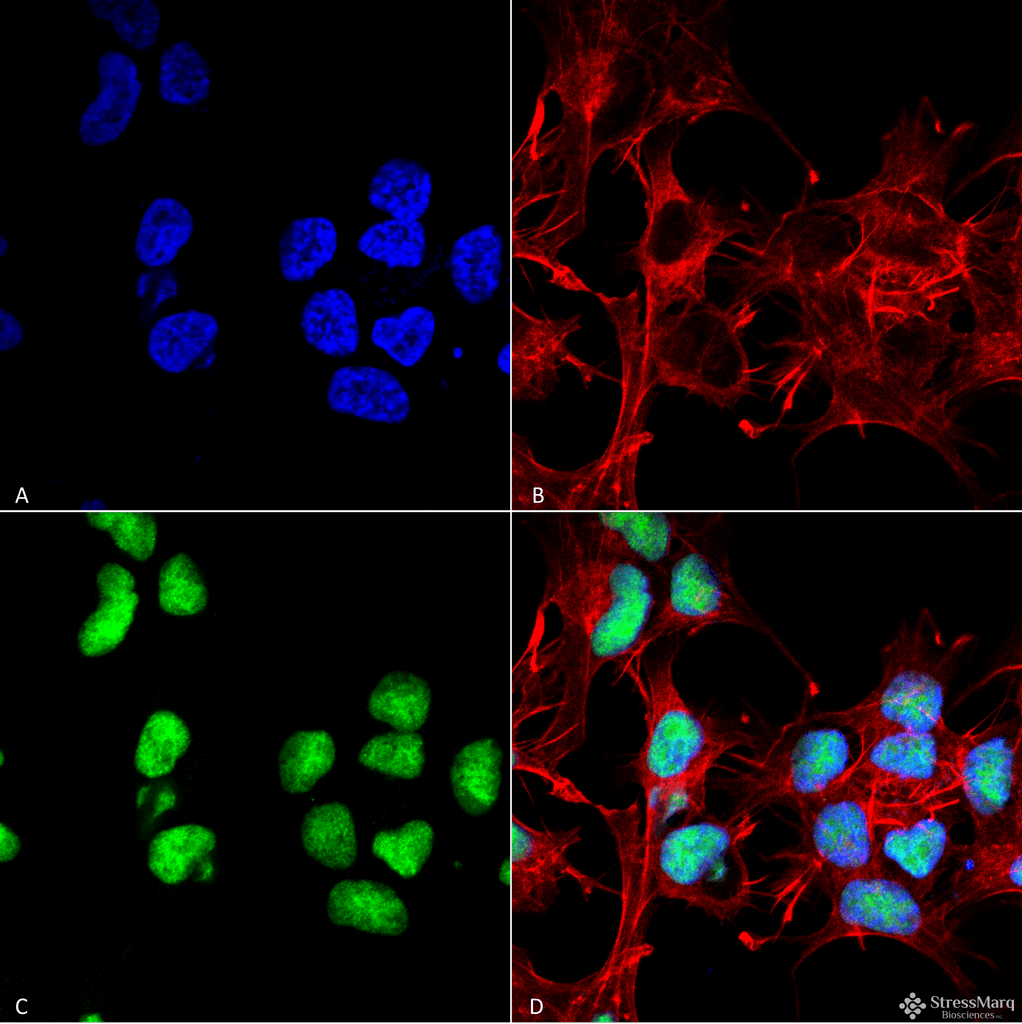 Immunocytochemistry/Immunofluorescence analysis using Rabbit Anti-Choline Acetyltransferase Polyclonal Antibody (56571). Tissue: Neuroblastoma cell line (SK-N-BE). Species: Human. Fixation: 4% Formaldehyde for 15 min at RT. Primary Antibody: Rabbit Anti-Choline Acetyltransferase Polyclonal Antibody (56571) at 1:100 for 60 min at RT. Secondary Antibody: Goat Anti-Rabbit ATTO 488 at 1:100 for 60 min at RT. Counterstain: Phalloidin Texas Red F-Actin stain; DAPI (blue) nuclear stain at 1:1000, 1:5000 for 60min RT, 5min RT. Localization: Nucleus. Magnification: 60X. (A) DAPI (blue) nuclear stain (B) Phalloidin Texas Red F-Actin stain (C) Choline Acetyltransferase Antibody (D) Composite.
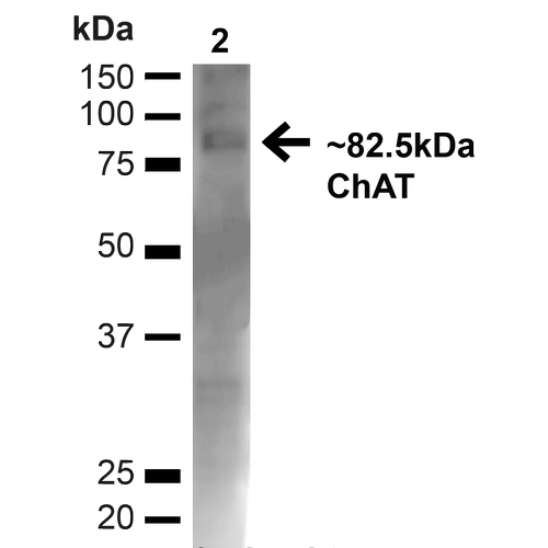 Western blot analysis of Mouse Brain showing detection of ~82.5kDa Choline Acetyltransferase protein using Rabbit Anti-Choline Acetyltransferase Polyclonal Antibody (56571). Lane 1: MW Ladder. Lane 2: Mouse Brain (20 µg). Load: 20 µg. Block: 5% milk + TBST for 1 hour at RT. Primary Antibody: Rabbit Anti-Choline Acetyltransferase Polyclonal Antibody (56571) at 1:1000 for 1 hour at RT. Secondary Antibody: Goat Anti-Rabbit: HRP at 1:2000 for 1 hour at RT. Color Development: TMB solution for 12 min at RT. Predicted/Observed Size: ~82.5kDa.
 - -
- -
Antibody DetailsProduct DetailsReactive Species Human ⋅ Mouse Host Species Rabbit Immunogen Synthetic peptide corresponding to amino acids from the N-terminus to the mid-protein of human choline o-acetyltransferase. Product Concentration 1.0 mg/ml Formulation PBS, pH 7.4, 50% glycerol, 0.09% sodium azide. State of Matter Liquid Product Preparation Purified by peptide immuno-affinity chromatography Storage and Handling This antibody is stable for at least one (1) year at -20°C. Regulatory Status For in vitro investigational use only. Not intended for therapeutic or diagnostic applications. Country of Origin USA Shipping Next Day 2-8°C Applications and Recommended Usage? Quality Tested by Leinco Immunoblotting: use at dilution of 1:1,000. A band of ~80kDa is detected.
Immunofluorescence: use at dilution of 1:100.Detection of choline acetyltransferase in mouse brain lysate with #56571 diluted 1:1,000 These are recommended working dilutions. Endusers should determine optimal dilutions for their applications. Each investigator should determine their own optimal working dilution for specific applications. See directions on lot specific datasheets, as information may periodically change. DescriptionDescriptionSpecificity This antibody recognizes human and mouse choline acetyltransferase. Background Choline acetyltransferase (ChAT) catalyzes the transfer of an acetyl group from acetyl-CoA to choline to produce acetylcholine. ChAT is found in high concentration in the central nervous system and peripheral nervous system. Alterations in concentrations of acetylcholine and ChAT have been associated with Alzheimer disease and Amyotrophic Lateral Sclerosis (ALS). Function Catalyzes the reversible synthesis of acetylcholine (ACh) from acetyl CoA and choline at cholinergic synapses. {PubMed:17144655}. NCBI Gene Bank ID UniProt.org Research Area Neuroscience References & CitationsTechnical ProtocolsICC IF  Certificate of Analysis |
 Products are for research use only. Not for use in diagnostic or therapeutic procedures.