

Anti-GIT1 Antibody (56537)


Anti-GIT1 Antibody (56537)
Product No.: 56537
- -
- -
Clone S39B-8 Target GIT1 Formats AvailableView All Product Type Monoclonal Isotype Mouse IgG1 Applications ICC , IF , IP , WB |
Data
 Immunocytochemistry/Immunofluorescence analysis using Mouse Anti-GIT1 Monoclonal Antibody, Clone S39B-8 (56537). Tissue: Neuroblastoma cells (SH-SY5Y). Species: Human. Fixation: 4% PFA for 15 min. Primary Antibody: Mouse Anti-GIT1 Monoclonal Antibody (56537) at 1:50 for overnight at 4°C with slow rocking. Secondary Antibody: AlexaFluor 488 at 1:1000 for 1 hour at RT. Counterstain: Phalloidin-iFluor 647 (red) F-Actin stain; Hoechst (blue) nuclear stain at 1:800, 1.6mM for 20 min at RT. (A) Hoechst (blue) nuclear stain. (B) Phalloidin-iFluor 647 (red) F-Actin stain. (C) GIT1 Antibody (D) Composite.
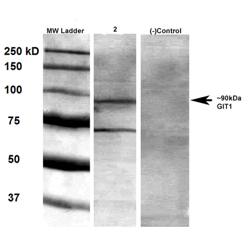 Western Blot analysis of Rat brain membrane lysate showing detection of GIT1 protein using Mouse Anti-GIT1 Monoclonal Antibody, Clone S39B-8 (56537). Primary Antibody: Mouse Anti-GIT1 Monoclonal Antibody (56537) at 1:1000.
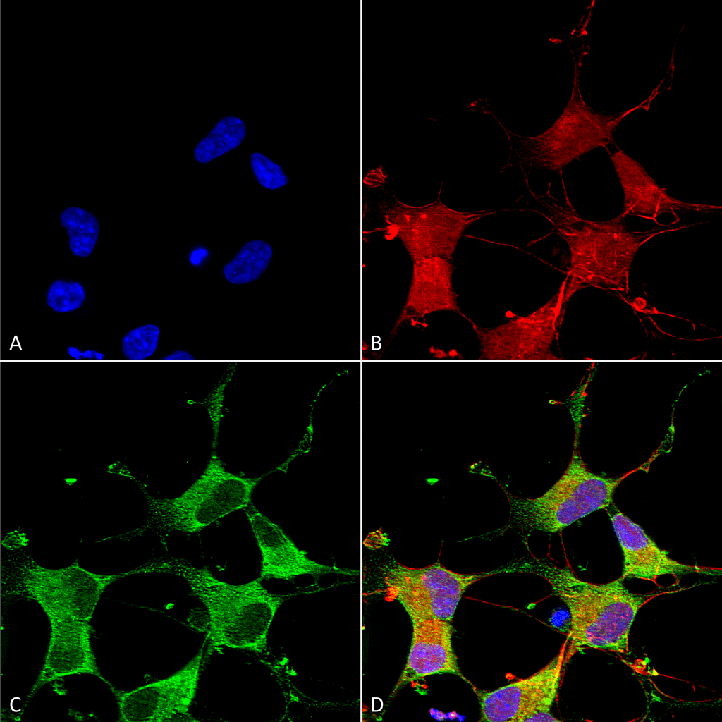
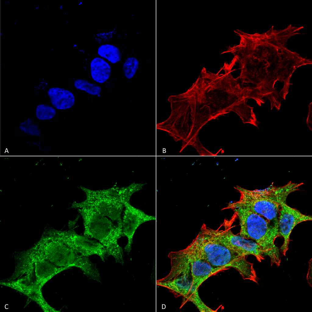 Immunocytochemistry/Immunofluorescence analysis using Mouse Anti-GIT1 Monoclonal Antibody, Clone S39B-8 (56537). Tissue: Neuroblastoma cell line (SK-N-BE). Species: Human. Fixation: 4% Formaldehyde for 15 min at RT. Primary Antibody: Mouse Anti-GIT1 Monoclonal Antibody (56537) at 1:100 for 60 min at RT. Secondary Antibody: Goat Anti-Mouse ATTO 488 at 1:100 for 60 min at RT. Counterstain: Phalloidin Texas Red F-Actin stain; DAPI (blue) nuclear stain at 1:1000; 1:5000 for 60 min RT, 5 min RT. Localization: Cytoplasm . Magnification: 60X. (A) DAPI (blue) nuclear stain. (B) Phalloidin Texas Red F-Actin stain. (C) GIT1 Antibody. (D) Composite.
 - -
- -
Antibody DetailsProduct DetailsReactive Species Human ⋅ Mouse ⋅ Rat Host Species Mouse Immunogen Fusion protein corresponding to aa 375-770 (C-terminus) of rat GIT1 (accession no.Q9Z272). Product Concentration 1.0 mg/ml Formulation PBS, pH 7.4, 50% glycerol, 0.09% sodium azide.Purified by Protein G affinity chromatography. State of Matter Liquid Product Preparation Purified by Protein G affinity chromatography Storage and Handling This antibody is stable for at least one (1) year at -20°C. Avoid repeated freeze-thaw cycles.
Regulatory Status For in vitro investigational use only. Not
intended for diagnostic or therapeutic
applications. Country of Origin USA Shipping Next Day 2-8°C Applications and Recommended Usage? Quality Tested by Leinco Immunoblotting: use at 1ug/mL. Predicted molecular weight is ~90kDa.
Positive control: rat brain lysate. These are recommended concentrations. Endusers should determine optimal concentrations for their applications. Each investigator should determine their own optimal working dilution for specific applications. See directions on lot specific datasheets, as information may periodically change. DescriptionDescriptionSpecificity This antibody recognizes human, mouse
and rat GIT1. It does not cross-react
with GIT2. Background G-protein coupled receptor (GPCR) kinase interacting proteins 1 and 2 (GIT1 and GIT2) are highly conserved ubiquitous scaffold proteins involved in localized signaling to help regulate focal contact assembly and cytoskeletal dynamics. GIT proteins contain multiple interaction domains for small GTPases, kinases, the Rho family GEF PIX, and the focal adhesion protein paxillin. GIT1 is localized to focal adhesions, cyto- plasmic complexes and membrane protrusions, and it regulates cell protrusion formation and cell migration. NCBI Gene Bank ID UniProt.org Research Area Neuroscience References & CitationsTechnical ProtocolsICC IF   Certificate of Analysis |
Formats Available
 Products are for research use only. Not for use in diagnostic or therapeutic procedures.