

Anti-Human Caspase-14 (CT)


Data
- -
- -
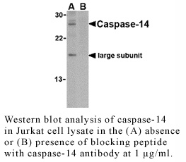
Antibody DetailsProduct DetailsReactive Species Human Host Species Rabbit Immunogen PN:C1287 Product Concentration 0.5 mg/ml Formulation This polyclonal antibody is formulated in phosphate buffered saline (PBS) pH 7.4 containing 0.02% sodium azide as a preservative. Storage and Handling This polyclonal antibody is stable for at least one week when stored at 2-8°C. For long term storage, aliquot in working volumes without diluting and store at –20°C in a manual defrost freezer. Avoid Repeated Freeze Thaw Cycles. Country of Origin USA Shipping Next Day Ambient RRIDAB_2828393 Each investigator should determine their own optimal working dilution for specific applications. See directions on lot specific datasheets, as information may periodically change. DescriptionDescriptionSpecificity Rabbit Anti-Human Caspase-14 recognizes an epitope near the C-terminus of mouse, rat and human Caspase-14. This polyclonal antibody was purified using affinity chromatography. Background Caspases are a family of cysteine proteases that can be divided into apoptotic and inflammatory caspase subfamilies. Unlike the apoptotic caspases, members of the inflammatory subfamily are generally not involved in cell death but are associated with the immune response to microbial pathogens. Members of this subfamily include caspase-1, -4, -5, and -12 and can activate proinflammatory cytokines such as IL-1b and IL-18. Caspase-14 is highly expressed in embryonic but not adult tissues. It is processed and activated by caspase 8 and caspase 10 in vitro, and by anti-Fas agonist antibody or TNF-related apoptosis inducing ligand in vivo. The expression and processing of this caspase may be involved in the keratinocyte terminal differentiation, which is important for the formation of the skin barrier. PubMed References & Citations1. Martinon, F. and Tschopp, J.(2004) Cell 117:561-74. 2. Zhivotovsky, B. and Orrenius, S. (2005) Biophys. Res. Comm. 331:859-67. 3. Flavell, RA. et al. (1995) Science 267:2000-3. 4. Gracie, JA. et al. (2003) J. Leukoc. Biol. 73:213-24. Technical Protocols Certificate of Analysis |
Related Products
 Products are for research use only. Not for use in diagnostic or therapeutic procedures.