

Anti-Human PD-L1 Antibody (34068)


Anti-Human PD-L1 Antibody (34068)
Product No.: 34068
- -
- -
| Clone G411.1 Target PD-L1 Formats AvailableView All Product Type Monoclonal Antibody Alternate Names PD-L1, PDCD1 ligand 1, Programmed death ligand 1, hPD-L1, B7 homolog 1, B7-H1, CD antigen CD274 Isotype Rabbit IgG Applications IHC |
- -
- -
| Select Product Size | Qty Max: Min: 1 Step: 1 $250.00 |
Data
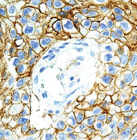 Immunohistochemistry: Detection of PD-L1 in human lung tumor with #34068 diluted 1:100-1:200.
- -
- -
Antibody DetailsProduct DetailsReactive Species Human Immunogen Recombinant human PD-L1. Formulation Tris buffer, pH 7.3-7.7, 1% BSA, 0.1% sodium azide. State of Matter Liquid Product Preparation Purified by immunoaffinity chromatography Storage and Handling Store at 2-8°C. Do not freeze. Shipping Next Day 2-8°C Applications and Recommended Usage? Quality Tested by Leinco Immunohistochemistry: use at a dilution of 1:100-1:200 on formalin-fixed, paraffin-embedded samples after heat-induced epitope retrieval at pH 9 for 10-30 minutes. Key IHC Advantages: - Optimized for Immunohistochemistry: Delivers robust, highly specific staining in formalin-fixed, paraffin-embedded (FFPE) human tissue sections. - Targeted Detection: Accurately visualizes PD-L1 (CD274/B7-H1) upregulation in the tumor microenvironment. - Reliable for Oncology Research: Ensures high-sensitivity, publication-ready IHC data for studies focused on cancer immune evasion and checkpoint inhibitor pathways. Each investigator should determine their own optimal working dilution for specific applications. See directions on lot specific datasheets, as information may periodically change. DescriptionDescriptionSpecificity Rabbit Polyclonal Antibody specific to PD-L1 Background Programmed Death-Ligand 1 (PD-L1 / CD274 / B7-H1) is a transmembrane protein that helps tumor cells evade the host immune system by binding to the PD-1 receptor. Detecting PD-L1 overexpression via immunohistochemistry (IHC) has become a cornerstone of modern oncology research and immunotherapy development. Our Anti-Human PD-L1 Antibody is expressly validated for high-resolution IHC staining on human tissue samples. Upregulation of PD-L1 is linked to increased tumor aggressiveness in renal cell carcinoma, and when assessed alongside CD8+ lymphocyte density, it acts as a key predictor in stage III non-small-cell lung cancer (NSCLC), breast cancer, and melanoma. Designed for reliable performance in FFPE tissues, this IHC-optimized antibody provides researchers with the high sensitivity and specificity needed to accurately map PD-L1 expression in the tumor microenvironment. Function Plays a critical role in induction and maintenance of immune tolerance to self (PubMed:11015443, PubMed:28813417, PubMed:28813410). As a ligand for the inhibitory receptor PDCD1/PD-1, modulates the activation threshold of T-cells and limits T-cell effector response (PubMed:11015443, PubMed:28813417, PubMed:28813410). Through a yet unknown activating receptor, may costimulate T-cell subsets that predominantly produce interleukin-10 (IL10) (PubMed:10581077). {PubMed:10581077, PubMed:11015443, PubMed:28813410, PubMed:28813417}.; The PDCD1-mediated inhibitory pathway is exploited by tumors to attenuate anti-tumor immunity and escape destruction by the immune system, thereby facilitating tumor survival (PubMed:28813417, PubMed:28813410). The interaction with PDCD1/PD-1 inhibits cytotoxic T lymphocytes (CTLs) effector function (By similarity). The blockage of the PDCD1-mediated pathway results in the reversal of the exhausted T-cell phenotype and the normalization of the anti-tumor response, providing a rationale for cancer immunotherapy (By similarity). {UniProtKB:Q9EP73, PubMed:28813410, PubMed:28813417}. NCBI Gene Bank ID UniProt.org Research Area Cancer Research References & Citations |
Formats Available
- -
- -
Prod No. | Description |
|---|---|
| 34068 |
 Products are for research use only. Not for use in diagnostic or therapeutic procedures.