

Anti-Insulin Receptor (α subunit) [Clone 83-14]


Anti-Insulin Receptor (α subunit) [Clone 83-14]
Product No.: 50101
- -
- -
Clone 83-14 Target Insulin receptor (α subunit) Formats AvailableView All Product Type Monoclonal Alternate Names IR, EC 2.7.10.1, CD antigen CD220 [Cleaved into: Insulin receptor subunitα; Insulin receptor subunit β] Isotype Mouse IgG2a Applications Act , IP , WB , Inhibition , Stimulation |
Data
- -
- -
Antibody DetailsProduct DetailsReactive Species Human Host Species Mouse Immunogen IM-9 lymphocytes Product Concentration Lot Specific Formulation PBS, pH 7.4. State of Matter Liquid Product Preparation This antibody is manufactured in an animal free facility using only in vitro protein free cell culture techniques and are purified by a multi-step process including the use of protein A or G to assure extremely low levels of endotoxins, leachable protein A or aggregates. Storage and Handling This antibody is stable for at least one (1) year at -20°C to -70°C. Store product in appropriate aliquots to avoid multiple freeze-thaw cycles. Country of Origin USA Shipping Next Day 2-8°C Each investigator should determine their own optimal working dilution for specific applications. See directions on lot specific datasheets, as information may periodically change. DescriptionDescriptionSpecificity Mouse Monoclonal Antibody specific to human insulin receptor; low-level cross-reactivity with bovine
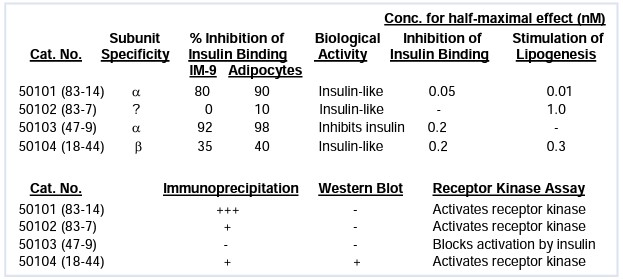
and rabbit insulin receptor. Background Anti-Insulin Receptor (α-subunit) [Clone 83-14] Overview of the Insulin Receptor (INSR / CD220) The Insulin Receptor (INSR), also known as CD220, is a high-affinity receptor tyrosine kinase essential for regulating glucose homeostasis and lipid metabolism. It exists as a heterotetramer consisting of two extracellular alpha (α) subunits and two transmembrane beta (β) subunits. Upon insulin binding to the α-subunit, the receptor undergoes a conformational change that activates the intrinsic kinase activity of the β-subunits, triggering the PI3K/Akt and MAPK signaling pathways. Specificity and Epitope Mapping of Clone 83-14 Clone 83-14 is a well-characterized mouse monoclonal antibody (IgG2a) that specifically recognizes the extracellular alpha subunit of the human insulin receptor. Technical mapping has localized its binding epitope to amino acids 469–592 (encoded by exons 7 and 8). Unlike many other clones, 83-14 is highly specific for the human receptor and shows no cross-reactivity with the rat insulin receptor or the closely related human Type 1 IGF receptor (IGF-1R), making it an ideal tool for clean, targeted research1. Functional Utility: Inhibition and Stimulation One of the most valuable characteristics of Clone 83-14 is its dual-action functional capability. It is widely utilized in scientific literature as a: - Potent Inhibitor: It can block insulin binding by approximately 80%, serving as a powerful tool for studies on receptor blocking and competitive binding assays. - Insulin-like Agonist: In specific carrier-free formulations, Clone 83-14 can act as a surrogate agonist, triggering receptor phosphorylation and downstream signaling even in the absence of insulin. This makes it highly relevant for research into insulin resistance syndromes and mutant receptor activation. Applications in Research & Drug Delivery Beyond standard validated applications like Western Blot (WB) and Immunoprecipitation (IP), Clone 83-14 has gained significant attention in Blood-Brain Barrier (BBB) research. It is recognized as one of the most effective transport vectors for delivering pharmaceutical payloads across the BBB via receptor-mediated transcytosis. Whether you are investigating Type 2 Diabetes (T2D), metabolic disorders, or oncogenic signaling, Leinco’s Clone 83-14 provides the high purity and specificity required for reproducible results. Function Receptor tyrosine kinase which mediates the pleiotropic actions of insulin. Binding of insulin leads to phosphorylation of several intracellular substrates, including, insulin receptor substrates (IRS1, 2, 3, 4), SHC, GAB1, CBL and other signaling intermediates. Each of these phosphorylated proteins serve as docking proteins for other signaling proteins that contain Src-homology-2 domains (SH2 domain) that specifically rUniProtKB:P15208, PubMed:12138094, PubMed:16314505, PubMed:16831875, PubMed:8257688, PubMed:8276809, PubMed:8452530, PubMed:9428692}. NCBI Gene Bank ID UniProt.org Research Area Growth Factors, Cytokines, Receptors References & Citations1.) Journal of Biology Chemistry Volume 265, Issue 17, 15 June 1990, Pages 9970-9977 2.) Soos et al. (1986) Biochem J 235: 199. 3.) Brindle et al. (1990) Biochem J 268: 615. 4.) Prigent et al. (1990) J Biol Chem 265: 9970 Technical ProtocolsAct   Inhibition Stimulation Certificate of Analysis |
Related Products
- -
- -
Formats Available
 Products are for research use only. Not for use in diagnostic or therapeutic procedures.