

Anti-mGluR1/5 Glutamate Receptor Antibody (56545)


Anti-mGluR1/5 Glutamate Receptor Antibody (56545)
Product No.: 56545
- -
- -
Clone S75-3 Target mGluR1/5 Glutamate Receptor Formats AvailableView All Product Type Monoclonal Alternate Names mGluR5 Isotype Mouse IgG2a Applications ICC , IF , IHC , IP , WB |
Data
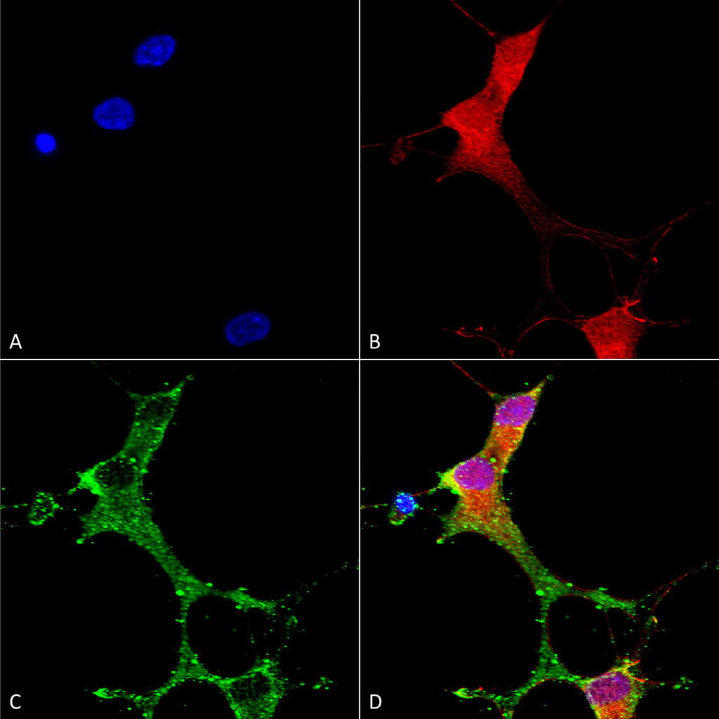 Immunocytochemistry/Immunofluorescence analysis using Mouse Anti-mGluR1/5 Monoclonal Antibody, Clone S75-3 (56545). Tissue: Neuroblastoma cells (SH-SY5Y). Species: Human. Fixation: 4% PFA for 15 min. Primary Antibody: Mouse Anti-mGluR1/5 Monoclonal Antibody (56545) at 1:50 for overnight at 4°C with slow rocking. Secondary Antibody: AlexaFluor 488 at 1:1000 for 1 hour at RT. Counterstain: Phalloidin-iFluor 647 (red) F-Actin stain; Hoechst (blue) nuclear stain at 1:800, 1.6mM for 20 min at RT. (A) Hoechst (blue) nuclear stain. (B) Phalloidin-iFluor 647 (red) F-Actin stain. (C) mGluR1/5 Antibody (D) Composite.
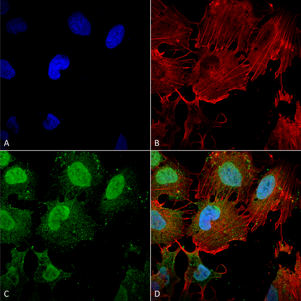 Immunocytochemistry/Immunofluorescence analysis using Mouse Anti-mGluR1/5 glutamate receptor Monoclonal Antibody, Clone S75-3 (56545). Tissue: Neuroblastoma cell line (SK-N-BE). Species: Human. Fixation: 4% Formaldehyde for 15 min at RT. Primary Antibody: Mouse Anti-mGluR1/5 glutamate receptor Monoclonal Antibody (56545) at 1:100 for 60 min at RT. Secondary Antibody: Goat Anti-Mouse ATTO 488 at 1:200 for 60 min at RT. Counterstain: Phalloidin Texas Red F-Actin stain; DAPI (blue) nuclear stain at 1:1000, 1:5000 for 60 min at RT, 5 min at RT. Localization: Cell Membrane, Cytoplasm, Nucleus. Magnification: 60X. (A) DAPI (blue) nuclear stain. (B) Phalloidin Texas Red F-Actin stain. (C) mGluR1/5 glutamate receptor Antibody. (D) Composite.
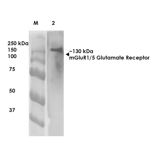 Western Blot analysis of Rat Brain Membrane showing detection of ~130 kDa mGluR5 protein using Mouse Anti-mGluR5 Monoclonal Antibody, Clone S75-33 (56545). Lane 1: Molecular Weight (MW) Ladder. Lane 2: Rat Brain Membrane. Load: 10 µg. Block: 5% milk. Primary Antibody: Mouse Anti-mGluR5 Monoclonal Antibody (56545) at 1:1000 for 1 hour at RT. Secondary Antibody: Goat Anti-Mouse IgG: HRP at 1:200 for 1 hour at RT. Color Development: TMB solution for 10 min at RT. Predicted/Observed Size: ~130 kDa.
 - -
- -
Antibody DetailsProduct DetailsReactive Species Human ⋅ Mouse ⋅ Rat Host Species Mouse Immunogen Fusion protein corresponding to aa 824-1203 (cytoplasmic C-terminus) of rat mGluR5b (accession no. P31424). Product Concentration Lot Specific Formulation PBS, pH 7.4; 50% glycerol, 0.09% sodium azide. Purified by Protein G affinity chromatography. State of Matter Liquid Product Preparation Purified by Protein G affinity chromatography Storage and Handling This antibody is stable for at least one (1) year at -20°C. Avoid repeated freezing
and thawing. Regulatory Status For in vitro investigational use only. Not for
use in therapeutic or diagnostic procedures. Country of Origin USA Shipping Next Day 2-8°C Applications and Recommended Usage? Quality Tested by Leinco Immunoblotting: use at 1ug/mL. A band of ~130kDa is detected. Smaller fragments due to proteolysis are detected as well.
Positive control: Rat brain lysate. These are recommended concentrations. User should determine optimal concentrations for their application. Each investigator should determine their own optimal working dilution for specific applications. See directions on lot specific datasheets, as information may periodically change. DescriptionDescriptionSpecificity This antibody recognizes human, mouse,
and rat mGluR1 and mGluR5. Background The metabotropic glutamate receptors (mGluRs) are active through an indirect metabotropic process. They are members of the group C family of G-protein-coupled receptors, or GPCRs. Like all glutamate receptors, mGluRs bind glutamate which functions as an excitatory neurotransmitter. Metabotropic glutamate receptors are not ion channels. Instead, they activate biochemical cascades, leading to the modification of other proteins. This can lead to changes in the synapse's excitability. Eight different types of mGluRs, designated mGluR1 to mGluR8, are divided into groups I, II, and III. Receptor types are grouped based on receptor structure and physiological activity. Function G-protein coupled receptor for glutamate. Ligand binding causes a conformation change that triggers signaling via guanine nucleotide-binding proteins (G proteins) and modulates the activity of down-stream effectors. Signaling activates a phosphatidylinositol-calcium sPubMed:1320017, PubMed:21795692}. NCBI Gene Bank ID UniProt.org Research Area Neuroscience References & CitationsTechnical ProtocolsICC IF    Certificate of Analysis |
Formats Available
 Products are for research use only. Not for use in diagnostic or therapeutic procedures.