

Anti-p95/Nibrin Antibody (56249)


Anti-p95/Nibrin Antibody (56249)
Product No.: 56249
- -
- -
Clone 1D7 Target p95/Nibrin Formats AvailableView All Product Type Monoclonal Alternate Names Cell cycle regulatory protein p95, Nijmegen breakage syndrome protein 1 Isotype Mouse IgG1 Applications ICC , IF , IP , WB |
Data
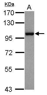 Sample (30 ug of whole cell lysate)
A: HepG2
7.5% SDS PAGE
56249 diluted at 1:1000
The HRP-conjugated anti-mouse IgG antibody was used to detect the primary antibody.
Sample (30 ug of whole cell lysate)
A: HepG2
7.5% SDS PAGE
56249 diluted at 1:1000
The HRP-conjugated anti-mouse IgG antibody was used to detect the primary antibody.![NBS1 antibody [1D7] detects NBS1 protein at nucleus by immunofluorescent analysis. Sample: A431 cells were fixed in 4% paraformaldehyde at RT for 15 min. Green: NBS1 stained by NBS1 antibody [1D7] (56249) diluted at 1:500. Red: phalloidin, a cytoskeleton marker, diluted at 1:200.](https://www.leinco.com/wp-content/uploads/2025/01/qed-bioscience-anti-p95-nibrin-antibody-56249-2.jpg) NBS1 antibody [1D7] detects NBS1 protein at nucleus by immunofluorescent analysis.
Sample: A431 cells were fixed in 4% paraformaldehyde at RT for 15 min.
Green: NBS1 stained by NBS1 antibody [1D7] (56249) diluted at 1:500.
Red: phalloidin, a cytoskeleton marker, diluted at 1:200.
NBS1 antibody [1D7] detects NBS1 protein at nucleus by immunofluorescent analysis.
Sample: A431 cells were fixed in 4% paraformaldehyde at RT for 15 min.
Green: NBS1 stained by NBS1 antibody [1D7] (56249) diluted at 1:500.
Red: phalloidin, a cytoskeleton marker, diluted at 1:200.![Various whole cell extracts (30 ug) were separated by 7.5% SDS-PAGE, and the membrane was blotted with NBS1 antibody [1D7] (56249) diluted at 1:500. The HRP-conjugated anti-mouse IgG antibody was used to detect the primary antibody.](https://www.leinco.com/wp-content/uploads/2025/01/qed-bioscience-anti-p95-nibrin-antibody-56249-3.jpg) Various whole cell extracts (30 ug) were separated by 7.5% SDS-PAGE, and the membrane was blotted with NBS1 antibody [1D7] (56249) diluted at 1:500. The HRP-conjugated anti-mouse IgG antibody was used to detect the primary antibody.
Various whole cell extracts (30 ug) were separated by 7.5% SDS-PAGE, and the membrane was blotted with NBS1 antibody [1D7] (56249) diluted at 1:500. The HRP-conjugated anti-mouse IgG antibody was used to detect the primary antibody. - -
- -
Antibody DetailsProduct DetailsReactive Species Human Host Species Mouse Immunogen Fusion protein containing the complete coding region of human p95/nibrin expressed in E. coli. Product Concentration Lot Specific Formulation PBS, pH 7.4. State of Matter Liquid Product Preparation Purified by Protein G affinity chromatography Storage and Handling This antibody is stable for at least one (1) year at -70°C. Avoid multiple freeze- thaw cycles. Regulatory Status Research Use Only Country of Origin USA Shipping Next Day 2-8°C Applications and Recommended Usage? Quality Tested by Leinco Immunoblotting,
Immunoprecipitation:: use at 1-2 ug/mL. In immunoblots, a band of 95 kD is detected. Positive controls: MCF-7, HeLa, or Raji cells. Each investigator should determine their own optimal working dilution for specific applications. See directions on lot specific datasheets, as information may periodically change. DescriptionSpecificity This antibody recognizes the 95 kD nibrin protein which contains a forkhead-associated domain that is adjacent to BRCT, a breast cancer C- terminal domain involved in protein- protein interactions. p95/nibrin is a member of the Mre11/Rad50 double- strand break repair complex. Function Component of the MRE11-RAD50-NBN (MRN complex) which plays a critical role in the cellular response to DNA damage and the maintenance of chromosome integrity. The complex is involved in double-strand break (DSB) repair, DNA rPubMed:10888888, PubMed:15616588, PubMed:19759395, PubMed:23762398, PubMed:26438602, PubMed:9705271}. NCBI Gene Bank ID UniProt.org Research Area Cancer Research References & CitationsTechnical ProtocolsICC IF   Certificate of Analysis |
Formats Available
 Products are for research use only. Not for use in diagnostic or therapeutic procedures.