

Anti-Shank 1 Antibody (56469)


Anti-Shank 1 Antibody (56469)
Product No.: 56469
- -
- -
Clone S22-21 Target Shank 1 Formats AvailableView All Product Type Monoclonal Alternate Names Shank1, GKAP/SAPAP-interacting protein, SPANK-1, Somatostatin receptor-interacting protein, SSTR-interacting protein, SSTRIP, Synamon Isotype Mouse IgG1 Applications IHC , WB , AM |
Data
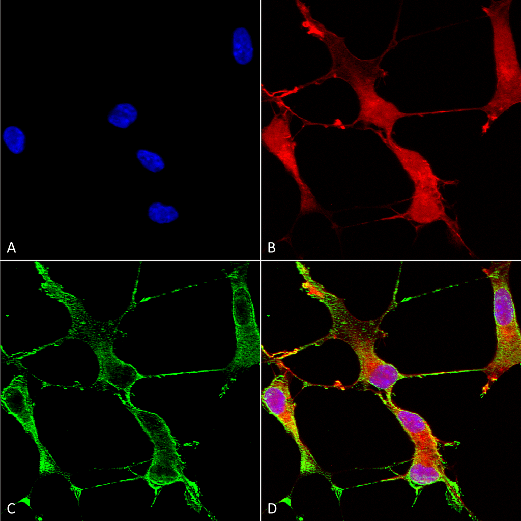 Immunocytochemistry/Immunofluorescence analysis using Mouse Anti-SHANK1 Monoclonal Antibody, Clone S22-21 (56469). Tissue: Neuroblastoma cells (SH-SY5Y). Species: Human. Fixation: 4% PFA for 15 min. Primary Antibody: Mouse Anti-SHANK1 Monoclonal Antibody (56469) at 1:50 for overnight at 4°C with slow rocking. Secondary Antibody: AlexaFluor 488 at 1:1000 for 1 hour at RT. Counterstain: Phalloidin-iFluor 647 (red) F-Actin stain; Hoechst (blue) nuclear stain at 1:800, 1.6mM for 20 min at RT. (A) Hoechst (blue) nuclear stain. (B) Phalloidin-iFluor 647 (red) F-Actin stain. (C) SHANK1 Antibody (D) Composite.
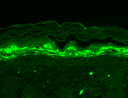 Immunohistochemistry analysis using Mouse Anti-SHANK1 Monoclonal Antibody, Clone S22-21 (56469). Tissue: backskin. Species: Mouse. Fixation: Bouin’s Fixative and paraffin-embedded. Primary Antibody: Mouse Anti-SHANK1 Monoclonal Antibody (56469) at 1:100 for 1 hour at RT. Secondary Antibody: FITC Goat Anti-Mouse (green) at 1:50 for 1 hour at RT. Localization: Filaggrin-like staining (upper layer aggregations of staining).
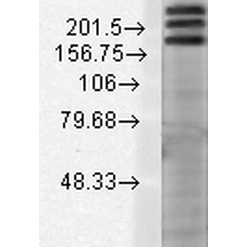 Western Blot analysis of Rat brain membrane lysate showing detection of SHANK1 protein using Mouse Anti-SHANK1 Monoclonal Antibody, Clone S22-21 (56469). Load: 15 µg. Block: 1.5% BSA for 30 minutes at RT. Primary Antibody: Mouse Anti-SHANK1 Monoclonal Antibody (56469) at 1:1000 for 2 hours at RT. Secondary Antibody: Sheep Anti-Mouse IgG: HRP for 1 hour at RT.
 - -
- -
Antibody DetailsProduct DetailsReactive Species Human ⋅ Mouse ⋅ Rat Host Species Mouse Immunogen Fusion protein corresponding to aa 469-691 (SH3/PDZ domains) of rat Shank1 (accession no. Q9WV48). This sequence is 96 Product Concentration Lot Specific Formulation PBS, pH 7.4; 50% glycerol, 0.09% sodium azide. State of Matter Liquid Product Preparation Purified by Protein G affinity chromatography Storage and Handling This antibody is stable for at least one (1) year at -20°C. Avoid repeated freezing
and thawing. Regulatory Status For in vitro investigational use only. Not for
use in therapeutic or diagnostic procedures. Country of Origin USA Shipping Next Day 2-8°C Applications and Recommended Usage? Quality Tested by Leinco Immunoblotting: use at 1-10ug/mL. Bands of ~190-220kDa (alternative splice variants) are detected.Immunohistochemistry and
Immunocytochemistry: use at 0.1-1ug/mL Immunofluorescence: use at 1-10ug/mL These are recommended concentrations. User should determine optimal concentrations for their application. Positive control: Adult rat brain. Each investigator should determine their own optimal working dilution for specific applications. See directions on lot specific datasheets, as information may periodically change. DescriptionDescriptionSpecificity This antibody recognizes human, mouse,
and rat Shank 1. It does not react with
Shank2 or 3. Background Shank proteins are a family of scaffold proteins identified through their interaction with a variety of membrane and cyto- plasmic proteins. Shank proteins at postsynaptic sites of excitatory synapses play roles in signal transmission into the postsynaptic neuron. Shank proteins are crucial in receptor tyrosine kinase signaling. Recent studies suggest that disruption of glutamate receptors at the Shank-postsynaptic platform could contribute to the destruction of post- synaptic density which underlies synaptic dysfunction and loss in Alzheimer's disease. Shank1 might be relevant to human autism spectrum disorders due to its differential role in specific cognitive processes along with its importance in synapse structure and function. Function Seems to be an adapter protein in the postsynaptic density (PSD) of excitatory synapses that interconnects receptors of the postsynaptic membrane including NMDA-type and metabotropic glutamate receptors, and the actin-based cytoskeleton. Plays a role in the structural and functional organization of the dendritic spine and synaptic junction. Overexpression promotes maturation of dendritic spines and the enlargement of spine heads via its ability to recruit Homer to postsynaptic sites, and enhances presynaptic function. {PubMed:11498055, PubMed:18287537}. NCBI Gene Bank ID UniProt.org Research Area Neuroscience References & CitationsTechnical Protocols  AM Certificate of Analysis |
Formats Available
 Products are for research use only. Not for use in diagnostic or therapeutic procedures.