

Anti-Shank 3 [Clone S69-46]


Anti-Shank 3 [Clone S69-46]
Product No.: 56463
- -
- -
Clone S69-46 Target Shank 3 Formats AvailableView All Product Type Monoclonal Alternate Names Shank3, Proline-rich synapse-associated protein 2, ProSAP2, SPANK-2 Isotype Mouse IgG2b Applications ICC , IF , IHC , IP , WB , AM |
Data
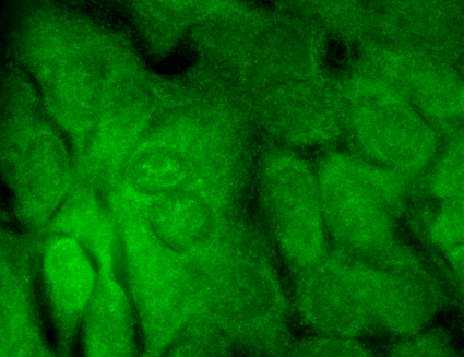 Immunocytochemistry/Immunofluorescence analysis using Mouse Anti-SHANK3 Monoclonal Antibody, Clone S69-46 (56463). Tissue: HaCaT cells. Species: Human. Fixation: Cold 100% methanol for 10 minutes at -20°C. Primary Antibody: Mouse Anti-SHANK3 Monoclonal Antibody (56463) at 1:100 for 1 hour at RT. Secondary Antibody: FITC Goat Anti-Mouse (green) at 1:50 for 1 hour at RT. Localization: Borderline positive.
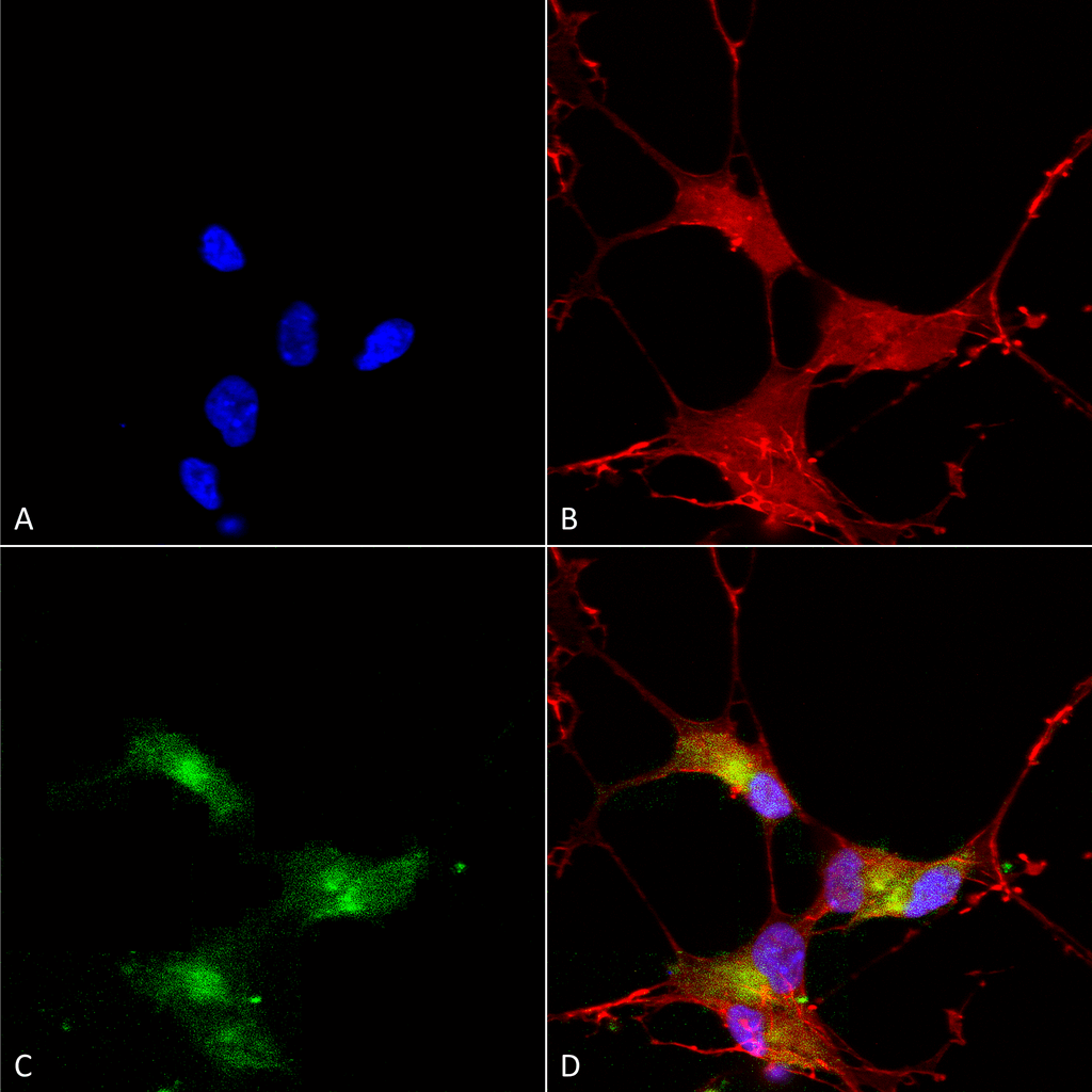 Immunocytochemistry/Immunofluorescence analysis using Mouse Anti-SHANK3 Monoclonal Antibody, Clone S69-46 (56463). Tissue: Neuroblastoma cells (SH-SY5Y). Species: Human. Fixation: 4% PFA for 15 min. Primary Antibody: Mouse Anti-SHANK3 Monoclonal Antibody (56463) at 1:50 for overnight at 4°C with slow rocking. Secondary Antibody: AlexaFluor 488 at 1:1000 for 1 hour at RT. Counterstain: Phalloidin-iFluor 647 (red) F-Actin stain; Hoechst (blue) nuclear stain at 1:800, 1.6mM for 20 min at RT. (A) Hoechst (blue) nuclear stain. (B) Phalloidin-iFluor 647 (red) F-Actin stain. (C) SHANK3 Antibody (D) Composite.
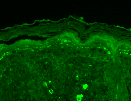 Immunohistochemistry analysis using Mouse Anti-SHANK3 Monoclonal Antibody, Clone S69-46 (56463). Tissue: backskin. Species: Mouse. Fixation: Bouin’s Fixative and paraffin-embedded. Primary Antibody: Mouse Anti-SHANK3 Monoclonal Antibody (56463) at 1:100 for 1 hour at RT. Secondary Antibody: FITC Goat Anti-Mouse (green) at 1:50 for 1 hour at RT. Localization: Early stages of filaggrin-like and dermal staining.
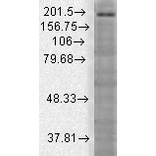 Western Blot analysis of Rat brain membrane lysate showing detection of SHANK3 protein using Mouse Anti-SHANK3 Monoclonal Antibody, Clone S69-46 (56463). Load: 15 µg. Block: 1.5% BSA for 30 minutes at RT. Primary Antibody: Mouse Anti-SHANK3 Monoclonal Antibody (56463) at 1:1000 for 2 hours at RT. Secondary Antibody: Sheep Anti-Mouse IgG: HRP for 1 hour at RT.
 - -
- -
Antibody DetailsProduct DetailsReactive Species Human ⋅ Mouse ⋅ Rat Host Species Mouse Immunogen Synthetic peptide corresponding to aa 840-857 (PEKLPGSLRKGIPRTKSV) of rat Shank3 (accession no. Q9JLU4). Product Concentration Lot Specific Formulation PBS, pH 7.4; 50% glycerol, 0.09% sodium azide. State of Matter Liquid Product Preparation Purified by Protein G affinity chromatography Storage and Handling This antibody is stable for at least one (1) year at -20°C. Avoid repeated freezing
and thawing. Regulatory Status For in vitro investigational use only. Not for
use in therapeutic or diagnostic procedures. Country of Origin USA Shipping Next Day 2-8°C Applications and Recommended Usage? Quality Tested by Leinco Immunoblotting: use at 1-10ug/mL. A band of ~40kDa is detected.Immunohistochemistry and
Immunocytochemistry: use at 0.1-1ug/mL Immunofluorescence: use at 1-10ug/mL These are recommended concentrations. User should determine optimal concentrations for their application. Positive control: Adult rat brain or lysate of COS cells transiently expressing Shank3. Each investigator should determine their own optimal working dilution for specific applications. See directions on lot specific datasheets, as information may periodically change. DescriptionDescriptionSpecificity This antibody recognizes human, mouse,
and rat Shank3. It does not cross-react
with Shank1 or Shank2. Background Shank proteins are a family of scaffold proteins identified through their interaction with a variety of membrane and cytoplasmic proteins. Shank proteins at postsynaptic sites of excitatory synapses play roles in signal transmission into the postsynaptic neuron. Shank proteins are crucial in receptor tyrosine kinase signaling; specifically, Shank3 can mediate Erk-MAPK and P13K signaling which is crucial for tubule formation. Shank 3 is also one of the latest genes to be associated with autism. A mutation in a single copy of Shank3 on chromosome 22q13 can result in communication disorders. Function Major scaffold postsynaptic density protein which interacts with multiple proteins and complexes to orchestrate the dendritic spine and synapse formation, maturation and maintenance. Interconnects receptors of the postsynaptic membrane including NMDA-type and metabotropic glutamate receptors via complexes with GKAP/PSD-95 and HOMER, respectively, and the actin-based cytoskeleton. Plays a role in the structural and functional organization of the dendritic spine and synaptic junction through the interaction with Arp2/3 and WAVE1 complex as well as the promotion of the F-actin clusters. By way of this control of actin dynamics, participates in the regulation of developing neurons growth cone motility and the NMDA receptor-signaling. Also modulates GRIA1 exocytosis and GRM5/MGLUR5 expression and signaling to control the AMPA and metabotropic glutamate receptor-mediated synaptic transmission and plasticity. May be required at an early stage of synapse formation and be inhibited by IGF1 to promote synapse maturation. {PubMed:21606927, PubMed:21795692, PubMed:23100419, PubMed:23897824, PubMed:24089484}. NCBI Gene Bank ID UniProt.org Research Area Neuroscience References & CitationsTechnical ProtocolsICC IF    AM Certificate of Analysis |
Formats Available
 Products are for research use only. Not for use in diagnostic or therapeutic procedures.