

Anti-TASK1 Potassium Channel Antibody (11578)

Anti-TASK1 Potassium Channel Antibody (11578)
Product No.: 11578
- -
- -
Clone S374-48 Target TASK1 Potassium Channel Formats AvailableView All Product Type Monoclonal Alternate Names Acid-sensitive potassium channel protein TASK-1, TWIK-related acid-sensitive K(+ channel 1, Two pore potassium channel KT3.1, Two pore K(+ channel KT3.1 Isotype Mouse IgG2b Applications ICC , IF , IHC , WB |
Data
 Immunocytochemistry/Immunofluorescence analysis using Mouse Anti-TASK1 Potassium Channel Monoclonal Antibody, Clone S374-48 (11578). Tissue: Neuroblastoma cells (SH-SY5Y). Species: Human. Fixation: 4% PFA for 15 min. Primary Antibody: Mouse Anti-TASK1 Potassium Channel Monoclonal Antibody (11578) at 1:50 for overnight at 4°C with slow rocking. Secondary Antibody: AlexaFluor 488 at 1:1000 for 1 hour at RT. Counterstain: Phalloidin-iFluor 647 (red) F-Actin stain; Hoechst (blue) nuclear stain at 1:800, 1.6mM for 20 min at RT. (A) Hoechst (blue) nuclear stain. (B) Phalloidin-iFluor 647 (red) F-Actin stain. (C) TASK1 Potassium Channel Antibody (D) Composite.
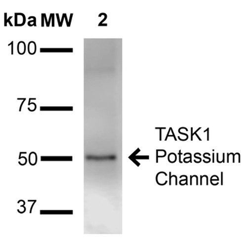 Western Blot analysis of Rat Brain Membrane showing detection of ~50 kDa TASK1 Potassium Channel protein using Mouse Anti-TASK1 Potassium Channel Monoclonal Antibody, Clone S374-48 (11578). Lane 1: Molecular Weight Ladder (MW). Lane 2: Rat brain membrane. Load: 15 µg. Block: 2% BSA and 2% Skim Milk in 1X TBST. Primary Antibody: Mouse Anti-TASK1 Potassium Channel Monoclonal Antibody (11578) at 1:1000 for 16 hours at 4°C. Secondary Antibody: Goat Anti-Mouse IgG: HRP at 1:2000 for 60 min at RT. Color Development: ECL solution for 6 min at RT. Predicted/Observed Size: ~50 kDa.
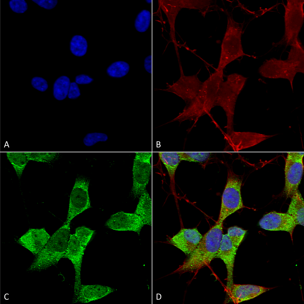
 Immunocytochemistry/Immunofluorescence analysis using Mouse Anti-TASK1 Potassium Channel Monoclonal Antibody, Clone S374-48 (11578). Tissue: Neuroblastoma cell line (SK-N-BE). Species: Human. Fixation: 4% Formaldehyde for 15 min at RT. Primary Antibody: Mouse Anti-TASK1 Potassium Channel Monoclonal Antibody (11578) at 1:100 for 60 min at RT. Secondary Antibody: Goat Anti-Mouse ATTO 488 at 1:100 for 60 min at RT. Counterstain: Phalloidin Texas Red F-Actin stain; DAPI (blue) nuclear stain at 1:1000, 1:5000 for 60min RT, 5min RT. Localization: Membrane. Magnification: 60X. (A) DAPI (blue) nuclear stain. (B) Phalloidin Texas Red F-Actin stain. (C) TASK1 Potassium Channel Antibody. (D) Composite.
 - -
- -
Antibody DetailsProduct DetailsReactivity Species Human ⋅ Mouse ⋅ Rat Host Species Mouse Immunogen Fusion protein corresponding to aa 251-411 (cytoplasmic C-terminus) of rat TASK1. This sequence is 96 Product Concentration 1.0 mg/ml Formulation PBS, pH 7.4, 0.1% sodium azide, 50% glycerol. State of Matter Liquid Product Preparation Purified by Protein G affinity chromatography Storage and Handling This product is stable for at least one (1) year at -20°C. Regulatory Status For in vitro investigational use only. Not for use in therapeutic or diagnostic procedures. Country of Origin USA Shipping Next Day 2-8°C Applications and Recommended Usage? Quality Tested by Leinco Immunoblotting: use at 1-5ug/mL. A band of ~50kDa is detected.
Immunofluorescence: use at 10ug/mL. These are recommended concentrations. Endusers should determine optimal concentrations for their application. Each investigator should determine their own optimal working dilution for specific applications. See directions on lot specific datasheets, as information may periodically change. DescriptionSpecificity This antibody recognizes human, mouse, and rat TASK1. Background TASK1 channels are members of the two-pore domain family of potassium channels whose structure consists of two pore-forming regions flanked by four membrane-spanning domains. The activity of these channels is sensitive to changes in extracellular pH in the physiological range. Like other two-pore domain family members, these channels show little time or voltage dependence. Thus they have characteristics of leak K+ channels, generating background currents that contribute to membrane potential and the shaping of cell excitability. Antigen DetailsFunction pH-dependent, voltage-insensitive, background potassium channel protein. Rectification direction results from potassium ion concentration on either side of the membrane. Acts as an outward rectifier when external potassium concentration is low. When external potassium concentration is high, current is inward. {UniProtKB:O14649}. NCBI Gene Bank ID UniProt.org Research Area Ion Channels References & CitationsTechnical ProtocolsICC IF   |
