

Recombinant Human PD-L1 (B7-H1)


Data
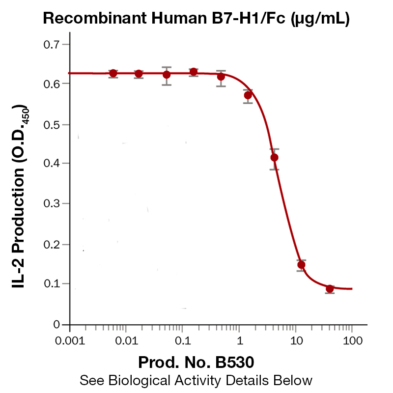
BackgroundPD-1 is a 50-55 kD member of the B7 Ig superfamily. PD-1 is also a member of the extended CD28/CTLA-4 family of T cell regulators and is suspected to play a role in lymphocyte clonal selection and peripheral tolerance. The ligands of PD-1 are PD-L1 and PD-L2, and are also members of the B7 Ig superfamily. PD-1 and its ligands negatively regulate immune responses. PD-L1, or B7-Homolog 1, is a 40 kD type I transmembrane protein that has been reported to costimulate T cell growth and cytokine production. The interaction of PD-1 with its ligand PD-L1 is critical in the inhibition of T cell responses that include T cell proliferation and cytokine production. PD-L1 has increased expression in several cancers. Inhibition of the interaction between PD-1 and PD-L1 can serve as an immune checkpoint blockade by improving T-cell responses In vitro and mediating preclinical antitumor activity. Within the field of checkpoint inhibition, combination therapy using anti-PD1 in conjunction with anti-CTLA4 has significant therapeutic potential for tumor treatments. PD-L2 is a 25 kD type I transmembrane ligand of PD-1. Via PD-1, PD-L2 can serve as a coinhibitor of T cell functions. Regulation of T cell responses, including enhanced T cell proliferation and cytokine production, can result from mAbs that block the PD-L2 and PD-1 interaction. Protein DetailsPurity >90% by SDS-PAGE and analyzed by silver stain. Endotoxin Level <0.01EU/µg as determined by the LAL method Biological Activity The biological activity of Human B7-H1 was determined by its ability to compete with biotin labeled in rhB7-H1 for the binding of rmPD-1/Fc in a functional ELISA. Fusion Protein Tag Fc Fusion Protein Protein Accession No. Amino Acid Sequence ft vtvpkdlyvv eygsnmtiec kfpvekqldl aalivyweme dkniiqfvhg eedlkvqhss yrqrarllkd qlslgnaalq itdvklqdag vyrcmisygg adykritvkv napynkinqr ilvvdpvtse heltcqaegy pkaeviwtss dhqvlsgktt ttnskreekl fnvtstlrin tttneifyct frrldpeenh taelvipelp lahppnertd iegrmdpksc dkthtcppcp apellggpsv flfppkpkdt lmisrtpevt cvvvdvshed pevkfnwyvd gvevhnaktk preeqynsty rvvsvltvlh qdwlngkeyk ckvsnkalpa piektiskak gqprepqvyt lppsrdeltk nqvsltclvk gfypsdiave wesngqpenn ykttppvlds dgsfflyskl tvdksrwqqg nvfscsvmhe alhnhytqks lslspgk
N-terminal Sequence Analysis Phe19 State of Matter Lyophilized Predicted Molecular Mass The predicted molecular weight of Recombinant Human B7-H1 is Mr 52 kDa. However, the actual molecular weight as observed by migration on SDS-PAGE is Mr 70-75 kDa. Predicted Molecular Mass 52 Formulation This recombinant protein was 0.2 µm filtered and lyophilized from modified Dulbecco’s phosphate buffered saline (1X PBS) and 300 mM NaCl, pH 7.2 – 7.4 with no calcium, magnesium, or preservatives. Storage and Stability This lyophilized protein is stable for six to twelve months when stored desiccated at -20°C to -70°C. After aseptic reconstitution, this protein may be stored at 2°C to 8°C for one month or at -20°C to -70°C in a manual defrost freezer. Avoid Repeated Freeze Thaw Cycles. See Product Insert for exact lot specific storage instructions. Country of Origin USA Shipping Next Day Ambient NCBI Gene Bank Leinco Protein AdvisorPowered by AI: AI is experimental and still learning how to provide the best assistance. It may occasionally generate incorrect or incomplete responses. Please do not rely solely on its recommendations when making purchasing decisions or designing experiments. Recombinant Human PD-L1 (B7-H1) is a valuable tool for a wide range of research applications, particularly in immunology and cancer biology. Here are several key reasons why you should consider using Recombinant Human PD-L1 in your research: 1. Study of Immune Checkpoint PathwaysPD-L1 (B7-H1) is a critical immune checkpoint molecule that interacts with PD-1 on T cells to regulate immune responses. Using recombinant PD-L1 allows you to:
2. Functional Assays and BioassaysRecombinant PD-L1 is suitable for various functional assays, including:
3. Development and Testing of TherapeuticsRecombinant PD-L1 is essential for:
4. Immune Tolerance and Autoimmunity ResearchPD-L1 plays a crucial role in maintaining immune tolerance and preventing autoimmunity. Using recombinant PD-L1 enables you to:
5. Cancer ImmunotherapyPD-L1 is highly expressed in many human cancers and is a key target for cancer immunotherapy. Recombinant PD-L1 can be used to:
6. High Purity and BioactivityRecombinant Human PD-L1 is typically produced with high purity and guaranteed bioactivity, ensuring reliable and reproducible results in your experiments. This is crucial for:
7. Versatility in ApplicationsRecombinant PD-L1 can be used in a variety of applications, including:
8. Support for Preclinical and Clinical ResearchRecombinant PD-L1 is widely used in preclinical studies to validate therapeutic targets and in clinical research to develop biomarkers for predicting response to immunotherapy. In summary, Recombinant Human PD-L1 (B7-H1) is an indispensable reagent for researchers studying immune checkpoint pathways, developing new immunotherapies, and exploring the complex interplay between the immune system and cancer. Its high purity, bioactivity, and versatility make it a reliable choice for a broad range of experimental applications. Yes, recombinant human PD-L1 (B7-H1) can be used as a standard for quantification or calibration in ELISA assays, provided it is of high purity, properly quantified, and compatible with your assay’s antibody pair and detection system. Key considerations and supporting details:
Limitations and best practices:
In summary, recombinant human PD-L1 is widely used and accepted as a standard for ELISA quantification, provided it is properly validated and compatible with your assay system. Recombinant Human PD-L1/B7-H1 proteins have been validated for a diverse range of applications in published research, reflecting their importance in immunological and cancer biology studies. Primary Research ApplicationsBinding and Protein-Protein Interaction Studies Recombinant PD-L1 proteins are extensively used in binding assays to characterize interactions with their natural ligands and engineered binding partners. These applications include screening high-affinity monoclonal antibodies against PD-L1 by ELISA and competition-based assays to identify anti-PD-1 antibodies. Biotinylated variants have been specifically optimized for biopanning experiments and other binding validation studies. Bioassay and Functional Assessment Functional bioassays represent a major application category, where recombinant PD-L1 is used to evaluate immunomodulatory effects. Notably, the bioactivity of PD-L1 Fc chimera proteins has been validated through their ability to inhibit anti-CD3 antibody-induced IL-2 secretion in human T lymphocyte primary cells, with reported effective concentrations (ED50) in the range of 0.075-0.75 μg/mL. Cell-Based Applications Recombinant PD-L1 has been validated for applications involving stem cell and immune cell maintenance or differentiation, as well as cell adhesion studies. Cell proliferation assays represent another validated application, where PD-L1's immunoregulatory effects on T cell proliferation can be measured. Immunological Detection and Analysis Surface plasmon resonance has been employed to characterize PD-L1 interactions in real-time kinetic studies. Additionally, recombinant PD-L1 serves as a control reagent for SDS-PAGE analysis and protein characterization. Therapeutic Development ContextBeyond basic research, recombinant PD-L1 proteins have supported the development of bifunctional fusion proteins targeting both PD-L1 and transforming growth factor-beta for anticancer immunotherapy, and have been instrumental in characterizing bispecific antibodies that simultaneously engage PD-L1 and other immune checkpoint pathways. To reconstitute and prepare Recombinant Human PD-L1 (B7-H1) protein for cell culture experiments, dissolve the lyophilized protein in sterile PBS at a concentration of 100 μg/mL for Fc-chimera formats, or as specified by the manufacturer for other formats. For His-tagged versions, reconstitution at 500 μg/mL in PBS is recommended. Always use sterile technique and avoid repeated freeze-thaw cycles. Step-by-step protocol:
Additional notes for cell culture experiments:
Summary Table:
Always refer to the specific product datasheet for any format-specific instructions. If the datasheet recommends sterile water instead of PBS, use sterile water. For cell culture, ensure all solutions are sterile and endotoxin-free. References & Citations1. Sheppard, KA. et al. (2004) FEBS Letters 574:37 2. Flies, DB. et al. (2007) J. Immunol. 30:251 3. Yamazaki, T. et al. (2002) J. Immunol. 169:5538 4. Thompson, RH. et al. (2004) Proc. Natl. Acad. Sci. USA 101:17174 Certificate of AnalysisIMPORTANT Use lot specific datasheet for all technical information pertaining to this recombinant protein. |
Related Products
Prod No. | Description |
|---|---|
P370 | |
P369 | |
P368 | |
P363 | |
P371 | |
B557 | |
B526 | |
B530 | |
B605 |
 Products are for research use only. Not for use in diagnostic or therapeutic procedures.