PD-1 is a 50-55 kD member of the B7 Ig superfamily. PD-1 is also a member of the extended CD28/CTLA-4 family of T cell regulators and is suspected to play a role in lymphocyte clonal selection and peripheral tolerance. The ligands of PD-1 are PD-L1 and PD-L2, and are also members of the B7 Ig superfamily. PD-1 and its ligands negatively regulate immune responses. PD-L1, or B7-Homolog 1, is a 40 kD type I transmembrane protein that has been reported to costimulate T cell growth and cytokine production. The interaction of PD-1 with its ligand PD-L1 is critical in the inhibition of T cell responses that include T cell proliferation and cytokine production. PD-L1 has increased expression in several cancers. Inhibition of the interaction between PD-1 and PD-L1 can serve as an immune checkpoint blockade by improving T-cell responses In vitro and mediating preclinical antitumor activity. Within the field of checkpoint inhibition, combination therapy using anti-PD1 in conjunction with anti-CTLA4 has significant therapeutic potential for tumor treatments. PD-L2 is a 25 kD type I transmembrane ligand of PD-1. Via PD-1, PD-L2 can serve as a coinhibitor of T cell functions. Regulation of T cell responses, including enhanced T cell proliferation and cytokine production, can result from mAbs that block the PD-L2 and PD-1 interaction.
Protein Details
Purity
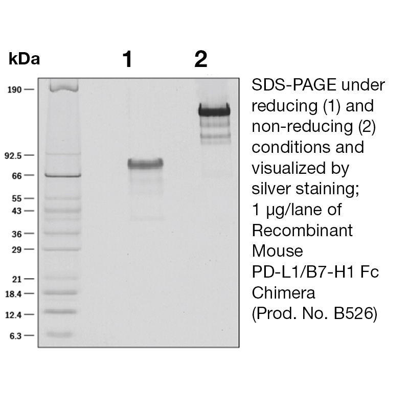
>90% by SDS-PAGE and analyzed by silver stain.
Endotoxin Level
<0.1 EU/µg as determined by the LAL method
Biological Activity
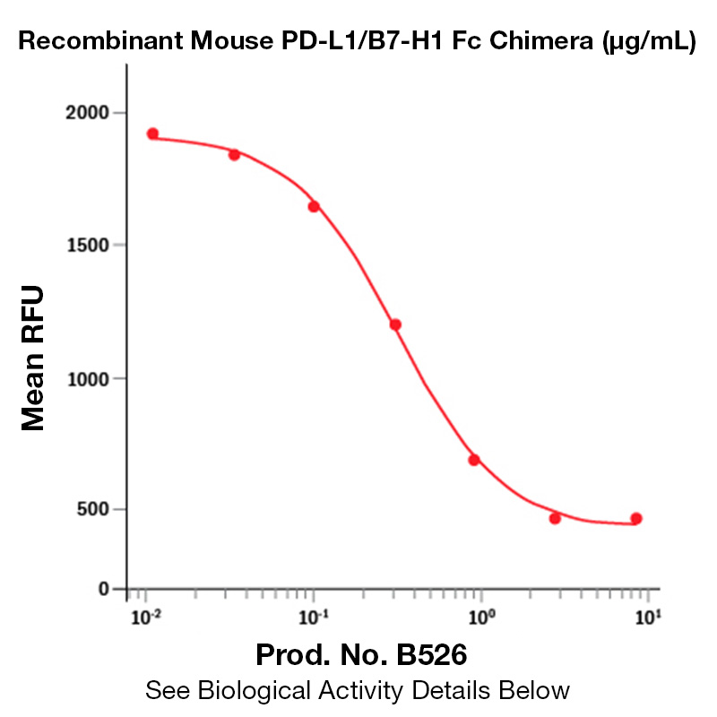
The biological activity of Mouse B7-H1 was determined by its ability to inhibit anti-CD3 induced proliferation of 72 hour PHA T cell blast. The expected ED<sub>50</sub> for this effect is typically 2.5 - 10 μg/mL.
The predicted molecular weight of Recombinant Mouse B7-H1 is Mr 51.3 kDa. However, the actual molecular weight as observed by migration on SDS-PAGE is Mr 75-85 kDa.
Predicted Molecular Mass
51.3
Formulation
This recombinant protein was 0.2 µm filtered and lyophilized from modified Dulbecco’s phosphate buffered saline (1X PBS) pH 7.2 – 7.4 with no calcium, magnesium, or preservatives.
Storage and Stability
This lyophilized protein is stable for six to twelve months when stored desiccated at -20°C to -70°C. After aseptic reconstitution, this protein may be stored at 2°C to 8°C for one month or at -20°C to -70°C in a manual defrost freezer. Avoid Repeated Freeze Thaw Cycles. See Product Insert for exact lot specific storage instructions.
Powered by AI: AI is experimental and still learning how to provide the best assistance. It may occasionally generate incorrect or incomplete responses. Please do not rely solely on its recommendations when making purchasing decisions or designing experiments.
Recombinant Mouse PD-L1 (B7-H1) is widely used in research applications to study immune checkpoint regulation, tumor immunology, and T cell biology due to its critical role as a ligand for PD-1, a key inhibitory receptor on immune cells.
Key scientific reasons to use recombinant mouse PD-L1 (B7-H1):
Functional studies of immune checkpoint pathways: PD-L1 binds to PD-1 on T cells, inhibiting their proliferation and cytokine production, which is central to immune tolerance and tumor immune evasion. Recombinant PD-L1 enables precise in vitro and in vivo modeling of these interactions.
Cancer immunotherapy research: PD-L1 is highly expressed on many murine and human cancers, contributing to immune escape. Recombinant PD-L1 is essential for evaluating checkpoint blockade strategies, such as monoclonal antibodies or engineered T cells, and for screening compounds that disrupt PD-1/PD-L1 signaling.
Bioassays and binding studies: Recombinant mouse PD-L1 is validated for binding to mouse PD-1 in a dose-dependent manner, making it suitable for ELISA, flow cytometry, and other assays to quantify or block PD-1/PD-L1 interactions.
Immunological tolerance and autoimmunity models: PD-L1 is involved in maintaining peripheral tolerance and preventing autoimmunity. Recombinant PD-L1 can be used to study regulatory T cell function, dendritic cell modulation, and the breakdown of tolerance in disease models.
Cell signaling and cytokine modulation: PD-L1 influences the production of anti-inflammatory cytokines (e.g., IL-10, IL-22) and inhibits pro-inflammatory Th17 cell development, making recombinant PD-L1 valuable for dissecting immune cell signaling pathways.
Typical applications include:
In vitro T cell activation and suppression assays.
In vivo mouse models of cancer, autoimmunity, and transplantation.
Screening and validation of immune checkpoint inhibitors.
Mechanistic studies of immune cell signaling and cytokine production.
Best practices:
Use recombinant PD-L1 in controlled bioassays to ensure reproducibility and specificity.
Avoid repeated freeze/thaw cycles to maintain protein activity.
Confirm binding activity with functional assays, such as ELISA or flow cytometry.
In summary, recombinant mouse PD-L1 (B7-H1) is a critical reagent for dissecting immune checkpoint mechanisms, developing immunotherapies, and modeling immune regulation in murine systems.
Yes, recombinant Mouse PD-L1 (B7-H1) protein can be used as a standard for quantification or calibration in ELISA assays, provided it is properly validated and matched to your assay system.
Essential context and supporting details:
Intended Use: Recombinant Mouse PD-L1 is commonly supplied as a standard in commercial ELISA kits designed to quantify PD-L1 in biological samples. These standards are used to generate a calibration curve, allowing for the quantification of PD-L1 concentrations in unknown samples.
Preparation: The standard should be reconstituted and diluted according to the manufacturer’s instructions or your assay protocol. For example, a typical protocol involves preparing a series of 2-fold serial dilutions in reagent diluent to create a standard curve.
Validation: It is critical to ensure that the recombinant PD-L1 standard is compatible with the capture and detection antibodies used in your ELISA. The protein should be of high purity, properly folded, and biologically active, as confirmed by binding assays or other functional tests.
Carrier Protein: Some recombinant proteins are supplied carrier-free or with BSA. Carrier-free preparations are preferred for ELISA standards to avoid interference or background signal.
Matrix Effects: When quantifying PD-L1 in complex matrices (e.g., plasma, serum, cell lysates), ensure that the standard is diluted in a matrix similar to your samples to account for potential matrix effects.
Best practices:
Use the recombinant PD-L1 standard supplied or recommended for your specific ELISA kit, as these have been validated for use with the kit’s antibodies and buffers.
If using a recombinant PD-L1 protein from a different source, validate its performance in your assay by comparing its standard curve to that of a known, validated standard.
Always refer to the Certificate of Analysis (CoA) for lot-specific information on concentration and reconstitution.
Limitations:
Recombinant standards may differ from native PD-L1 in post-translational modifications, which could affect antibody recognition. Confirm that your antibodies detect both recombinant and native forms equivalently.
Not all recombinant PD-L1 proteins are validated for use as ELISA standards; some are intended for bioassays or functional studies only.
In summary, recombinant Mouse PD-L1 is suitable as an ELISA standard if it is validated for this purpose and compatible with your assay system. Always follow best practices for standard preparation and assay validation to ensure accurate quantification.
Recombinant Mouse PD-L1 (B7-H1) has been validated in published research for a range of applications, primarily focused on immunological assays and functional studies.
Key validated applications include:
In Vivo Functional Studies: Used to modulate immune responses in mouse models, such as inducing immune tolerance, suppressing T cell activation, and studying tumor immune evasion.
ELISA (Enzyme-Linked Immunosorbent Assay): Employed as a capture reagent or standard for quantifying PD-L1 or its interactions in serum and cell-based samples.
Flow Cytometry: Utilized to detect and quantify PD-L1 binding or expression on cells, and to assess the functional consequences of PD-L1/PD-1 interactions.
Bioassays: Applied in cell-based assays to study PD-L1’s biological activity, including its effects on T cell proliferation, cytokine production, and immune checkpoint signaling.
Protein-Protein Interaction Assays: Used in binding assays, such as surface plasmon resonance, to characterize the interaction between PD-L1 and its receptors (e.g., PD-1, B7-1/CD80).
Blocking/Neutralization Assays: Validated for use in blocking PD-L1/PD-1 interactions to study immune checkpoint inhibition and its effects on immune cell function.
Enzyme Assays: Used to assess the enzymatic or signaling consequences of PD-L1 engagement in various cell types.
Western Blot and Immunohistochemistry: While more commonly validated for anti-PD-L1 antibodies, recombinant PD-L1 protein can serve as a positive control or standard in these applications.
Supporting details from published research:
In vivo studies have demonstrated the use of recombinant mouse PD-L1 to induce immune tolerance, suppress T cell activation, and promote regulatory T cell development.
ELISA and flow cytometry applications include quantifying PD-L1 levels and analyzing its binding to PD-1 or B7-1 on immune cells.
Bioassays have been used to assess the functional impact of PD-L1 on T cell proliferation, cytokine secretion, and immune checkpoint signaling.
Surface plasmon resonance and other protein-protein interaction assays have validated the direct binding of PD-L1 to its receptors.
Summary Table:
Application Type
Example Use Case/Assay
Reference
In Vivo Functional Studies
Immune modulation, tumor models
ELISA
Quantification, binding studies
Flow Cytometry
Cell surface binding, functional analysis
Bioassay
T cell proliferation, cytokine production
Protein-Protein Interaction
Surface plasmon resonance, binding kinetics
Blocking/Neutralization
Immune checkpoint inhibition studies
Enzyme Assay
Signaling pathway analysis
Western Blot/IHC
Positive control, standard
These applications are supported by multiple peer-reviewed studies and product validation data, confirming the utility of recombinant mouse PD-L1 (B7-H1) in both basic and translational immunology research.
To reconstitute and prepare Recombinant Mouse PD-L1 (B7-H1) protein for cell culture experiments, dissolve the lyophilized protein in sterile buffer according to the recommended concentration and handling guidelines for your specific construct.
General Protocols:
Fc Chimera Format: Reconstitute the lyophilized protein at 100 μg/mL in sterile PBS. Gently mix until fully dissolved. If needed, centrifuge briefly to remove any particulates.
His-tag Format: Reconstitute at 400 μg/mL in sterile PBS, or at ≤0.2 mg/mL in sterile double-distilled water. Mix gently by inversion or vortexing until fully dissolved.
Alternative Recommendations: Some protocols suggest reconstituting at 0.1–1 mg/mL in filtered deionized water. Always check the product datasheet for specific buffer and concentration recommendations.
Preparation for Cell Culture:
Aliquoting: After reconstitution, aliquot the protein into sterile tubes to avoid repeated freeze-thaw cycles.
Storage: Store aliquots at –20°C or colder under sterile conditions. Avoid repeated freeze/thaw cycles to maintain protein integrity.
Carrier Proteins (Optional): When preparing working solutions, you may add 0.2–1% BSA or HSA to stabilize the protein, especially for low concentration applications.
Sterility: Ensure all buffers and water used are sterile and endotoxin-free, especially for cell culture applications.
Usage Notes:
For bioassays, working concentrations typically range from 0.1–0.5 μg/mL for functional studies.
Always verify the activity and purity of the reconstituted protein before use in sensitive cell culture experiments.
Summary Table:
Format
Reconstitution Buffer
Concentration
Storage
Fc Chimera
Sterile PBS
100 μg/mL
–20°C or colder
His-tag
Sterile PBS or ddH₂O
400 μg/mL or ≤0.2 mg/mL
–20°C or colder
General
Filtered deionized water
0.1–1 mg/mL
–20°C or colder
Best Practices:
Use freshly reconstituted protein or thawed aliquots for each experiment.
Avoid repeated freeze-thaw cycles.
Confirm the absence of endotoxin if using in sensitive immune cell assays.
If your recombinant PD-L1 protein has a unique tag or formulation, always follow the manufacturer’s datasheet for precise instructions.
References & Citations
1. Sheppard, KA. et al. (2004) FEBS Letters 574:37
2. Flies, DB. et al. (2007) J. Immunol. 30:251
3. Yamazaki, T. et al. (2002) J. Immunol. 169:5538
4. Thompson, RH. et al. (2004) Proc. Natl. Acad. Sci. USA101:17174


 Products are for research use only. Not for use in diagnostic or therapeutic procedures.