

Accelerated Optical Clearing of Organoids for Fluorescent Imaging
Aqueous, solvent-free, high-throughput, optical clearing method that prepares your samples in hours, not days
Maintaining the 3D organization of an organoid while permitting imaging throughout is critical to understand the spatial relationships present in these tissues. However, traditional clearing methods provide 3D imaging at the expense of shrinkage, swelling, quenched reporters, hazardous solvents, and multiday protocols. ADAPT-3D takes a different, gentler route that accelerates the process so organoids become transparent in minutes to a few hours while size, structure, and fluorescent signal are preserved, without ever leaving the original 96 well plate.
Topics covered include:
Background on organoids and 3D imaging:
An overview of traditional methods and ADAPT-3D:
A comparison of organic-solvent (iDISCO/DISCO), hydrogel (CLARITY), hyperhydration (CUBIC), and ADAPT-3D methods on morphology preservation, reporter retention, reagent toxicity, and sample throughput.
How ADAPT-3D increases throughput and accelerates your workflow:
A complete overview of the ADAPT-3D protocol and the benefits and rationale behind each step.
The Science in Brief
Transparency is a refractive-index problem
Organoids are, by design, three-dimensional and can range from a few hundred micrometers to several millimeters in size, depending on the modeled tissue and maturation stage. Their increasing size and structural complexity creates an imaging challenge where in uncleared organoids, heterogeneity of refractive index and absorption by endogenous components scatter and attenuate excited and emitted fluorescence, progressively degrading image quality with depth. Here, we describe the ADAPT-3D workflow, an integrated aqueous clearing ecosystem developed for scalable three-dimensional organoid imaging, in which fixation, decolorization, partial delipidation, immunolabeling, and refractive-index matching are chemically coordinated and executed end-to-end in a single 96-well U-bottom plate.
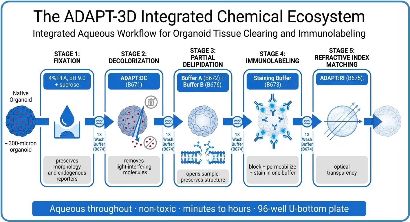
Figure 1. A single organoid passes through five chemically matched stages: fixation (4% PFA, pH 9.0 + sucrose), decolorization (ADAPT:DC, B671), optional partial delipidation (B672 + B676), immunolabeling (Staining Buffer, B673), and refractive-index matching (ADAPT:RI, B675). Each stage is bracketed by 1X Wash Buffer (B674) steps. Conceptual schematic from the white paper.
Why Leinco Reagents
The ADAPT-3D organoid clearing kit is designed to preserve spatial relationships while optimizing fluorescent readouts
Shrinkage distorts volume and inter-cellular spacing, swelling distorts network geometry, and reporter quenching erases the signal of interest. ADAPT-3D does not rely on toxic organic solvents and is designed to overcome these traditional limitations for organoids in the 100–500 µm size range while accelerating time to image acquisition.
| Attribute | CLARITY | iDISCO | CUBIC | ADAPT-3D |
|---|---|---|---|---|
| Processing time | Days–weeks | ~4 days | ~7 days | Minutes–hours |
| Morphology | Hydrogel swelling | Up to ~40% shrinkage | Swelling / tearing | No significant measurable change |
| Endogenous reporters | Variable | Diminished / lost | Diminished | Preserved (eGFP family, eYFP, tdTomato, CFP) |
| Chemistry | Acrylamide + detergents | Organic solvents | Aminoalcohol / urea | Aqueous, solvent-free |
| Format | Vials, manual transfer | Vials, manual transfer | Vials / multiwell | Single 96-well U-bottom plate |
Staining uses only 50 µL of buffer per well. That conserves antibody and lets you run a dilution series, secondary-only controls, and positive/negative controls in dedicated columns of the same plate. Transparency is maintained by immersion, so organoids are imaged in place in 100% ADAPT:RI. This keeps well registration fixed and eliminates mounting-induced distortion.
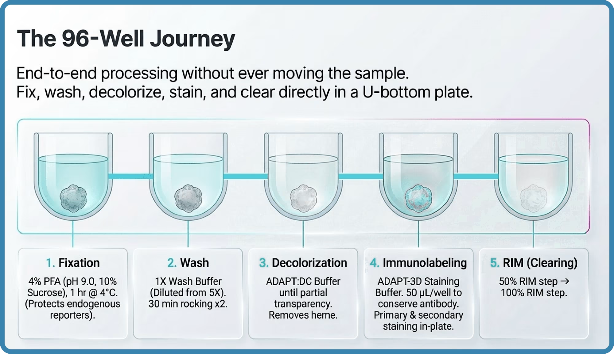
Figure 2. Fixation, washing, decolorization, immunolabeling, and two-step RI matching are all performed in a single 96-well U-bottom well, followed by in-plate imaging. Conceptual schematic from the white paper.
| The ADAPT-3D Core — Organoid Clearing Kit |
|---|
| ADAPT-3D Organoid Clearing Kit [A640] Covers 1 full 96-well plate through antibody labeling and clearing. |
| ADAPT-3D Decolorization Buffer, ADAPT:DC [B671] Removes light-interfering heme and pigments before labeling. |
| ADAPT-3D Immunostaining Buffer [B673] Blocks, permeabilizes, and carries primary and secondary antibodies in one buffer. |
| ADAPT-3D 5X Wash Buffer [B674] Removes reagents between steps. |
| ADAPT-3D Refractive Index Match Buffer, ADAPT:RI [B675] Two-step 50% → 100% protocol to a final RI of ≈1.50–1.51. |
| For organoids approaching ~500 µm, add Partial Delipidation Buffers A + B [B672, & B676] mixed 1:1. This step must run in glass, not plastic. |