

Anti-LAMP2 Antibody (56272)

Anti-LAMP2 Antibody (56272)
Product No.: 56272
- -
- -
Clone GL2A7 Target LAMP2 Formats AvailableView All Product Type Monoclonal Alternate Names LAMP-2, Lysosome-associated membrane protein 2, CD107 antigen-like family member B, Lysosomal membrane glycoprotein type B, LGP-B, CD antigen CD107b Isotype Rat IgG1 Applications ICC , IF , IP , WB |
Data
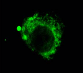 Immunocytochemistry/Immunofluorescence analysis using Rat Anti-LAMP2 Monoclonal Antibody, Clone GL2A7 (56272). Tissue: Corneal Endothelial Cell (CEC). Species: Rabbit. Primary Antibody: Rat Anti-LAMP2 Monoclonal Antibody (56272) at 1:1000. Secondary Antibody: FITC Goat Anti-Rat (green). Courtesy of: Eunduck E.P. Kay, Doheny Eye Institute.
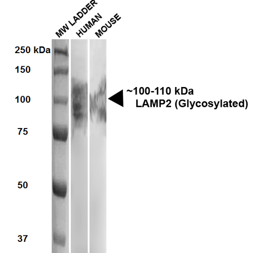 Western Blot analysis of Human, Mouse HEK293 and 3T3NIH cell lysates showing detection of ~100-110 kDa LAMP2 protein using Rat Anti-LAMP2 Monoclonal Antibody, Clone GL2A7 (56272). Lane 1: MW ladder. Lane 2: Human HEK293 lysate (20 µg). Lane 3: Mouse 3T3NIH lysate (10 µg). Block: 5% milk + TBST for 1 hour at RT. Primary Antibody: Rat Anti-LAMP2 Monoclonal Antibody (56272) at 1:500 for 1 hour at RT. Secondary Antibody: HRP Goat Anti-Rat at 1:100 for 1 hour at RT. Color Development: TMB solution for 5 min at RT. Predicted/Observed Size: ~100-110 kDa.
 - -
- -
Antibody DetailsProduct DetailsReactivity Species Mouse ⋅ Rabbit Host Species Rat Immunogen Gluteraldehyde-fixed mouse liver lysosomes Product Concentration Lot Specific Formulation PBS, pH 7.4. State of Matter Liquid Product Preparation Purified by Protein G affinity chromatography Storage and Handling This antibody is stable for at least one (1) year at -20°C.Avoid repeated freezing and thawing. Regulatory Status For in vitro investigational use only. Not for use in therapeutic or diagnostic procedures. Country of Origin USA Shipping Next Day 2-8°C Applications and Recommended Usage? Quality Tested by Leinco Immunoblotting: Use at 1-2ug/mL. A band of ~100-110kDa (glycosylated) is detected.
Immunofluorescence: Use at 1-2ug/mL. This antibody labels lysosomes and late endosomes in cells permeabilized with saponin. These are recommended concentrations. Enduser should determine optimal concentrations for their application. Each investigator should determine their own optimal working dilution for specific applications. See directions on lot specific datasheets, as information may periodically change. DescriptionSpecificity This antibody recognizes human, mouse, and rabbit LAMP2 (~100-110 kDa).Accession no.: NP_001017959.1 Gene ID: 16784 Background Lysosome-associated membrane proteins (LAMP1 and LAMP2) are major constituents of the lysosomal membrane. These two proteins have closely related structures with 37% sequence homology. Both are transmembrane glycoproteins localized primarily in lysosomes and late endosomes. LAMP2 has also been detected at the plasma membrane of cells undergoing differentiation and activation and in cells that secrete lysosomal hydrolases. Cell surface LAMP1 and LAMP2 promote adhesion of human peripheral blood mononuclear cells (PBMC) to vascular endothelium which suggests that the LAMP proteins are involved in adhesion of PBMC to sites of inflammation. Defects in LAMP2 are associated with Danon disease. Antigen DetailsFunction Plays an important role in chaperone-mediated autophagy, a process that mediates lysosomal degradation of proteins in response to various stresses and as part of the normal turnover of proteins with a long biological half-live (PubMed:10972293). Functions by binding target proteins, such as GAPDH and MLLT11, and targeting them for lysosomal degradation (By similarity). Required for the fusion of autophagosomes with lysosomes during autophagy (PubMed:27628032). Cells that lack LAMP2 express normal levels of VAMP8, but fail to accumulate STX17 on autophagosomes, which is the most likely explanation for the lack of fusion between autophagosomes and lysosomes (PubMed:27628032). Required for normal degradation of the contents of autophagosomes (PubMed:10972293, PubMed:12221139). Plays a role in lysosomal protein degradation in response to starvation (PubMed:27628032). Required for efficient MHCII-mediated presentation of exogenous antigens via its function in lysosomal protein degradation; antigenic peptides generated by proteases in the endosomal/lysosomal compartment are captured by nascent MHCII subunits. Is not required for efficient MHCII-mediated presentation of endogenous antigens (By similarity). {UniProtKB:P13473, UniProtKB:P17046, PubMed:10972293, PubMed:12221139, PubMed:27628032}. NCBI Gene Bank ID UniProt.org Research Area Cell Adhesion References & CitationsTechnical ProtocolsICC IF   |
