

Anti-Ankyrin-G Antibody (56519)

Anti-Ankyrin-G Antibody (56519)
Product No.: 56519
- -
- -
Clone S106-20 Target Ankyrin-G Formats AvailableView All Product Type Monoclonal Alternate Names ANK-3, Ankyrin-G Isotype Mouse IgG1 Applications ICC , IF , WB |
Data
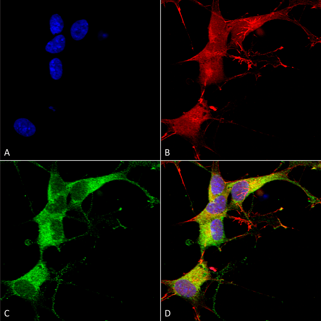 Immunocytochemistry/Immunofluorescence analysis using Mouse Anti-Ankyrin G Monoclonal Antibody, Clone S106-20 (56519). Tissue: Neuroblastoma cells (SH-SY5Y). Species: Human. Fixation: 4% PFA for 15 min. Primary Antibody: Mouse Anti-Ankyrin G Monoclonal Antibody (56519) at 1:100 for overnight at 4°C with slow rocking. Secondary Antibody: AlexaFluor 488 at 1:1000 for 1 hour at RT. Counterstain: Phalloidin-iFluor 647 (red) F-Actin stain; Hoechst (blue) nuclear stain at 1:800, 1.6mM for 20 min at RT. (A) Hoechst (blue) nuclear stain. (B) Phalloidin-iFluor 647 (red) F-Actin stain. (C) Ankyrin G Antibody (D) Composite.
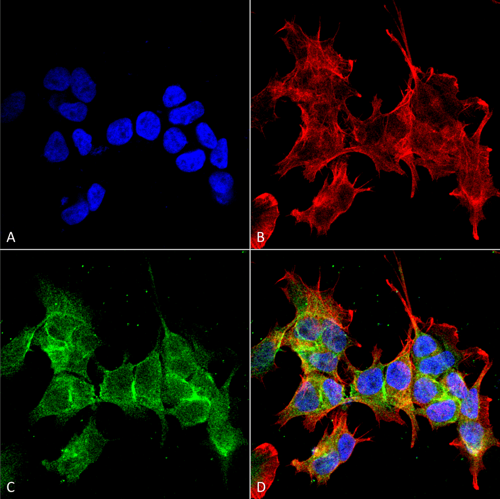 Immunocytochemistry/Immunofluorescence analysis using Mouse Anti-Ankyrin G Monoclonal Antibody, Clone S106-20 (56519). Tissue: Neuroblastoma cell line (SK-N-BE). Species: Human. Fixation: 4% Formaldehyde for 15 min at RT. Primary Antibody: Mouse Anti-Ankyrin G Monoclonal Antibody (56519) at 1:100 for 60 min at RT. Secondary Antibody: Goat Anti-Mouse ATTO 488 at 1:200 for 60 min at RT. Counterstain: Phalloidin Texas Red F-Actin stain; DAPI (blue) nuclear stain at 1:1000, 1:5000 for 60 min at RT, 5 min at RT. Localization: Cytoplasm . Magnification: 60X. (A) DAPI (blue) nuclear stain. (B) Phalloidin Texas Red F-Actin stain. (C) Ankyrin G Antibody. (D) Composite.
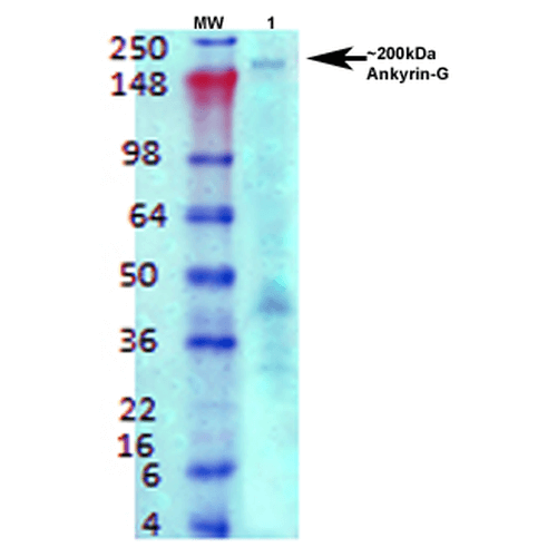 Western Blot analysis of Rat brain membrane lysate showing detection of Ankyrin G protein using Mouse Anti-Ankyrin G Monoclonal Antibody, Clone S106-20 (56519). Primary Antibody: Mouse Anti-Ankyrin G Monoclonal Antibody (56519) at 1:1000.
 - -
- -
Antibody DetailsProduct DetailsReactive Species Human ⋅ Mouse ⋅ Rat Host Species Mouse Immunogen Fusion protein corresponding to ~1,000 C-terminal amino acids of human Ankyrin G (accession no. NP_066267.2). Product Concentration 1.0 mg/ml Formulation PBS, pH 7.4, 50% glycerol, 0.09% sodium azide.Purified by Protein G affinity chromatography. State of Matter Liquid Product Preparation Purified by Protein G affinity chromatography Storage and Handling This product is stable for at least 1 year at -20°C. Freeze in multiple aliquots to avoid repeated freeze-thaw cycles. Regulatory Status For in vitro investigational use only. Not for
use in therapeutic or diagnostic procedures. Country of Origin USA Shipping Next Day 2-8°C Applications and Recommended Usage? Quality Tested by Leinco Immunoblotting: use at 1-2ug/mL. A band of >200kDa is detected.
These are recommended concentrations. Enduser should determine optimal concentrations for their applications. Positive control: rat brain membranes. Each investigator should determine their own optimal working dilution for specific applications. See directions on lot specific datasheets, as information may periodically change. DescriptionDescriptionSpecificity This antibody recognizes human, mouse,
and rat Ankyrin-G. Background Ankyrins are a family of adaptor proteins that mediate the attachment of integral membrane proteins to the spectrin-actin based membrane skeleton. Ankyrins have binding sites for the beta subunit of spectrin and for at least 12 families of integral membrane proteins. This linkage is required to maintain the integrity of the plasma membranes and to anchor specific ion channels, ion exchangers, and ion transporters in the plasma membrane. Function In skeletal muscle, required for costamere localization of DMD and betaDAG1 (By similarity). Membrane-cytoskeleton linker. May participate in the maintenance/targeting of ion channels and cell adhesion molecules at the nodes of Ranvier and axonal initial segments. Regulates KCNA1 channel activity in function of dietary Mg(2+) levels, and thereby contributes to the regulation of renal Mg(2+) reabsorption (PubMed:23903368). {PubMed:17974005}.; [Isoform 5]: May be part of a Golgi-specific membrane cytoskeleton in association with beta-spectrin. {PubMed:17974005}. NCBI Gene Bank ID UniProt.org Research Area Neuroscience References & CitationsTechnical ProtocolsICC IF  Certificate of Analysis |
