

Anti-Decoy Receptor 3 (DcR3) [Clone MD3E2] – Purified in vivo GOLD™ Functional Grade

Anti-Decoy Receptor 3 (DcR3) [Clone MD3E2] – Purified in vivo GOLD™ Functional Grade
Product No.: D114
- -
- -
Product No.D114 Clone MD3E2 Target DcR3 Product Type Recombinant Monoclonal Antibody Alternate Names TR6, M68 Isotype Mouse IgG Applications ELISA , FA , FACS , IHC , WB |
Data
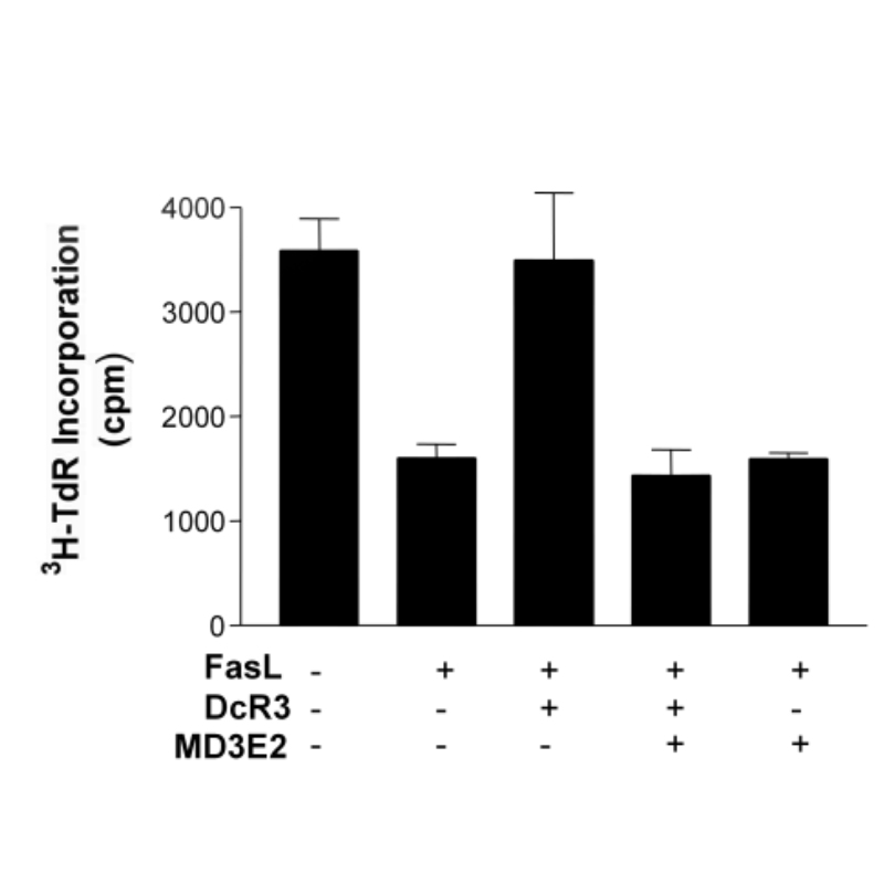 DcR3 inhibition of FasL-induced cancer cell death. A breast cancer cell line, MDA435, was seeded at 5000 cells/well in 96-well microtiter tissue culture plates overnight. Plated cells were treated with fresh cell culture media (150 μl/well) containing recombinant FasL (100 ng/ml) either in the presence or absence of soluble human recombinant DcR3 (500 ng/ml). DcR3 specificity was confirmed with 1 μg/ml of neutralizing mAb (MD3E2) or an iso-type matching (IgG1) control mAb. Cell proliferation was analyzed using 3H-TdR incorporation (cpm)
 - -
- -
Antibody DetailsProduct DetailsReactive Species Human Host Species Mouse Expression Host HEK-293 Cells Recommended Dilution Buffer Immunogen Full length human DcR3 Product Concentration ≥ 5.0 mg/ml Endotoxin Level < 1.0 EU/mg as determined by the LAL method Purity ≥95% monomer by analytical SEC ⋅ >95% by SDS Page Formulation This monoclonal antibody is aseptically packaged and formulated in 0.01 M phosphate buffered saline (150 mM NaCl) PBS pH 7.2 - 7.4 with no carrier protein, potassium, calcium or preservatives added. Due to inherent biochemical properties of antibodies, certain products may be prone to precipitation over time. Precipitation may be removed by aseptic centrifugation and/or filtration. State of Matter Liquid Product Preparation Functional grade preclinical antibodies are manufactured in an animal free facility using only in vitro protein free cell culture techniques and are purified by a multi-step process including the use of protein A or G to assure extremely low levels of endotoxins, leachable protein A or aggregates. Storage and Handling Functional grade preclinical antibodies may be stored sterile as received at 2-8°C for up to one month. For longer term storage, aseptically aliquot in working volumes without diluting and store at ≤ -70°C. Avoid Repeated Freeze Thaw Cycles. Regulatory Status Research Use Only Country of Origin USA Shipping 2 – 8° C Wet Ice Additional Applications Reported In Literature ? ELISA, FA, FACS, IHC, WB Each investigator should determine their own optimal working dilution for specific applications. See directions on lot specific datasheets, as information may periodically change. DescriptionDescriptionSpecificity MD3E2 activity is directed against DcR3. Background DcR3 (also known as TR6, M68, and TNFRSF6B) is a member of the tumor necrosis factor
receptor superfamily1,2. DcR3 acts as a soluble decoy receptor that can modulate the effect of
TNF surface receptors by competing for binding to the TNF family ligands Fas ligand, LIGHT,
and TL1A. DcR3 also plays a role in the immune system by modulating T cell responses as well
as differentiation and maturation of dendritic cells. Additionally, DcR3 likely plays a role in
keratinocyte differentiation and egg fertilization2. DcR3 is normally expressed at very low levels but is upregulated in tumors and inflammatory tissues2. Accordingly, DcR3 is regarded as a potential biomarker for cancer metastasis and as a predictor of inflammatory disease progression. In tumors, upregulation of DcR3 prevents apoptosis and promotes growth, invasion, and angiogenesis. Additionally, upregulation of DcR3 as part of the TL1A/death receptor 3 (DR3/TNFRSF25) axis is important to the pathogenesis of various inflammatory and autoimmune diseases including silicosis, bacterial infection, coronary artery disease, inflammatory bowel disease, osteoarthritis, and rheumatoid arthritis. DcR3 can also be upregulated by ultraviolet light, epidermal growth factor, and transforming growth factor (TGF)-α. MD3E2 was generated against full-length human DcR3 by hybridoma technology1. An antibody panel was screened for binding to both the full-length protein as well as the exon I portion of DcR3, spanning residues 1-424. MD3E2 was found to selectively recognize the full-length DcR3. MD3E2 was produced for use in ELISA assays to quantify soluble DcR3. Antigen Distribution DcR3 is expressed in a wide variety of normal human tissues as well as
various cancers. DcR3 is detectable in granulosa cells, endometrial cells, theca cells,
undifferentiated primary keratinocytes, monocytes, dendritic cells, synovial fluids, and
chondrocytes, among others. Ligand/Receptor FasL, LIGHT, and TL1A NCBI Gene Bank ID UniProt.org Research Area Apoptosis . Cell Biology . Immunology References & Citations1 Chen J, Zhang L, Kim S. J Immunol Methods. 285(1):63-70. 2004. 2 Hsieh SL, Lin WW. J Biomed Sci. 24(1):39. 2017. Technical Protocols FA FACS   Certificate of Analysis |
