Anti-Human Rhodopsin – Purified
Anti-Human Rhodopsin – Purified
Product No.: R127
- -
- -
Clone RET-P1 Target Rhodopsin Formats AvailableView All Product Type Monoclonal Antibody Alternate Names RHO, MGC138309, MGC138311, OPN2, RP4, Visual Purple, Opsin 2 Isotype IgG1 Applications IHC FFPE , IP , WB |
Data
- -
- -
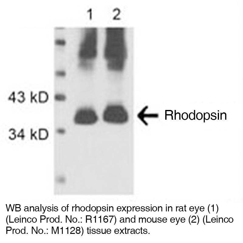
Antibody DetailsProduct DetailsReactive Species Human Host Species Mouse Product Concentration 0.5 mg/ml Formulation This purified antibody is formulated in 0.01 M phosphate buffered saline (150 mM NaCl) PBS pH 7.4, 1% BSA and 0.09% sodium azide as a preservative. Storage and Handling This purified antibody is stable when stored at 2-8°C. Do not freeze. Country of Origin USA Shipping Next Day Ambient RRIDAB_2831700 Each investigator should determine their own optimal working dilution for specific applications. See directions on lot specific datasheets, as information may periodically change. DescriptionDescriptionSpecificity Mouse Anti-Human Rhodopsin (Clone RET-P1) recognizes a (Mr 39 kD) glycoprotein which is a visual pigment in mammals, birds, amphibians and reptiles.1,2,3 Background Rhodopsin, also known as visual purple, is expressed in metazoan photoreceptor cells. It is a pigment of the retina that is responsible for both the formation of the photoreceptor cells and the first events in the perception of light. Rhodopsins belong to the class of G-protein coupled receptors. Rhodopsin is extremely sensitive to light, and enables night-vision. Exposed to white light, the pigment immediately bleaches, and it takes about 30 minutes to regenerate fully in humans. Antigen Distribution The Rhodopsin antigen is present on the photoreceptor cells in the retina. The antibody RET-P1 specifically labels the axons and synaptic pedicles of the rods being labelled.1,2,3 Research Area Cell Biology . Neuroscience References & Citations1. Barnstable, C. J. et al. (1980) Nature 286:231
2. Hicks, D. et al. (1987) J. Histochem. Cytochem. 35:1317
3. Treisman, J. E. et al. (1988) Molec. Cell. Biol. 8:1570 Technical ProtocolsCertificate of Analysis |


