

Anti-MDC1 [Clone P2B11]


Anti-MDC1 [Clone P2B11]
Product No.: 12596
- -
- -
Clone P2B11 Target MDC1 Formats AvailableView All Product Type Monoclonal Isotype Mouse IgG1 Applications ICC , IF , WB |
Data
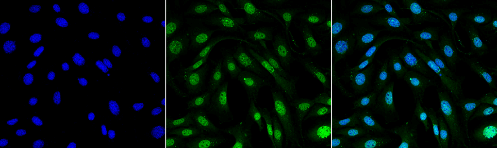 Immunocytochemistry/Immunofluorescence analysis using Mouse Anti-MDC1 Monoclonal Antibody, Clone P2B11 (12596). Tissue: Fibroblast cell line (NIH 3T3). Species: Mouse. Fixation: 4% Formaldehyde for 15 min at RT. Primary Antibody: Mouse Anti-MDC1 Monoclonal Antibody (12596) at 1:100 for 60 min at RT. Secondary Antibody: Goat Anti-Mouse ATTO 488 at 1:100 for 60 min at RT. Counterstain: DAPI (blue) nuclear stain at 1:5000 for 5 min RT. Localization: Nucleus. Magnification: 60X.
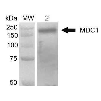 Western Blot analysis of Human Embryonic kidney epithelial cell line (HEK293T) lysate showing detection of 184 kDa MDC1 protein using Mouse Anti-MDC1 Monoclonal Antibody, Clone P2B11 (12596). Lane 1: MW ladder. Lane 2: 293Trap cell lysates. Load: 30 µg. Block: 5% Skim Milk in 1X TBST. Primary Antibody: Mouse Anti-MDC1 Monoclonal Antibody (12596) at 1:1000 for 2 hours RT. Secondary Antibody: Goat Anti-Mouse HRP: IgG at 1:2000 for 60 min at RT. Color Development: ECL solution for 5 min in RT. Predicted/Observed Size: 184 kDa.
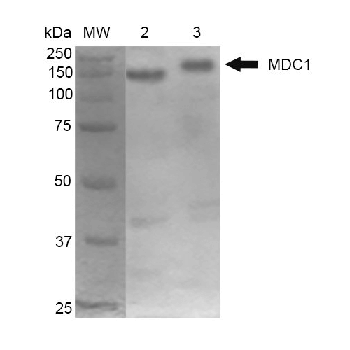 Western Blot analysis of Mouse Cortex and Cerebellum showing detection of 184 kDa MDC1 protein using Mouse Anti-MDC1 Monoclonal Antibody, Clone P2B11 (12596). Lane 1: MW ladder. Lane 2: Mouse Cortex. Lane 3: Mouse Cerebellum. Load: 10 µg. Block: 5% Skim Milk in 1X TBST. Primary Antibody: Mouse Anti-MDC1 Monoclonal Antibody (12596) at 1:1000 for 2 hours RT. Secondary Antibody: Goat Anti-Mouse at 1:2000 for 60 min at RT. Color Development: ECL solution for 5 min in RT. Predicted/Observed Size: 184 kDa.
 - -
- -
Antibody DetailsProduct DetailsReactivity Species Chimpanzee ⋅ Bovine ⋅ Human ⋅ Mouse Host Species Mouse Immunogen GST-tagged recombinant mouse MDC1 (accession no. NP_001010833.2). Product Concentration 1.0 mg/ml Formulation PBS, pH 7.4, 50% glycerol, 0.09% sodium azide. State of Matter Liquid Product Preparation Purified by Protein G affinity chromatography Storage and Handling This antibody is stable for at least one (1) year at -20°C. Avoid repeated freezethaw cycles. Regulatory Status For in vitro investigational use only. Not
intended for diagnostic or therapeutic
applications. Country of Origin USA Shipping Next Day 2-8°C Applications and Recommended Usage? Quality Tested by Leinco Immunocytochemistry Immunoblotting: use at 1ug/mL. A band of ~184kDa is detected. Note: There are 4 isoforms of human MDC1 with molecular weights of approximately 227, 198, 196, and 117kDa.(For more information see Lou et al. 2003, Nature 421: 957-961)
These are recommended concentrations; Enduser should determine optimal concentrations for their applications. Positive control: HeLa cell lysate. Each investigator should determine their own optimal working dilution for specific applications. See directions on lot specific datasheets, as information may periodically change. DescriptionSpecificity This antibody recognizes human, mouse, bovine and chimpanzee MDC1. Specific epitope is at the N-terminus of MDC1. Background Recent studies have shown that MDC1 (mediator of DNA damage checkpoint protein1) regulates many aspects of DNA damage response pathways, such as intra-S phase checkpoint, G2/M checkpoint, and radiation-induced apoptosis. Many proteins, such as ATM, BRCA1, and Chk2, interact with MDC1. MDC1 contains several protein-protein interaction domains. MDC1 appears to function as an adaptor protein, recruiting downstream proteins to upstream kinases and facilitating signal transduction following DNA damage. Antigen DetailsFunction Required for checkpoint mediated cell cycle arrest in response to DNA damage within both the S phase and G2/M phases of the cell cycle. May serve as a scaffold for the recruitment of DNA repair and signal transduction proteins to discrete foci of DNA damage marked by 'Ser-139' phosphorylation of histone H2AX. Also required for downstream events subsequent to the recruitment of these proteins. These include phosphorylation and activation of the ATM, CHEK1 and CHEK2 kinases, and stabilization of TP53 and apoptosis. ATM and CHEK2 may also be activated independently by a parallel pathway mediated by TP53BP1 (By similarity). {ECO:0000250}. NCBI Gene Bank ID UniProt.org Research Area DNA References & CitationsTechnical ProtocolsICC IF  |
Formats Available
 Products are for research use only. Not for use in diagnostic or therapeutic procedures.