

Anti-Nav1.8 Na+ Channel [S134]

Anti-Nav1.8 Na+ Channel [S134]
Product No.: 11523
- -
- -
Clone S134-12 Target Nav1.8 Na+ Channel Formats AvailableView All Product Type Monoclonal Alternate Names Peripheral nerve sodium channel 3, PN3, Sensory neuron sodium channel, Sodium channel protein type X subunitα, Voltage-gated sodium channel subunitα Nav1.8 Isotype Mouse IgG2a Applications ICC , IF , IHC , WB , AM |
Data
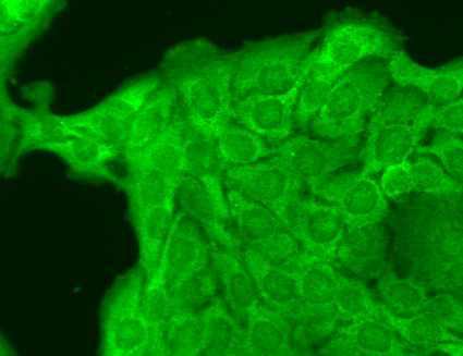 Immunocytochemistry/Immunofluorescence analysis using Mouse Anti-Nav1.8 Monoclonal Antibody, Clone S134 (11523). Tissue: HaCaT cells. Species: Human. Fixation: Cold 100% methanol for 10 minutes at -20°C. Primary Antibody: Mouse Anti-Nav1.8 Monoclonal Antibody (11523) at 1:100 for 1 hour at RT. Secondary Antibody: FITC Goat Anti-Mouse (green) at 1:50 for 1 hour at RT. Localization: Cytoplasmic staining and some dull nuclear staining.
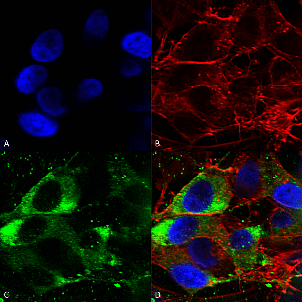 Immunocytochemistry/Immunofluorescence analysis using Mouse Anti-Nav1.8 Monoclonal Antibody, Clone S134 (11523). Tissue: Neuroblastoma cells (SH-SY5Y). Species: Human. Fixation: 4% PFA for 15 min. Primary Antibody: Mouse Anti-Nav1.8 Monoclonal Antibody (11523) at 1:50 for overnight at 4°C with slow rocking. Secondary Antibody: AlexaFluor 488 at 1:1000 for 1 hour at RT. Counterstain: Phalloidin-iFluor 647 (red) F-Actin stain; Hoechst (blue) nuclear stain at 1:800, 1.6mM for 20 min at RT. (A) Hoechst (blue) nuclear stain. (B) Phalloidin-iFluor 647 (red) F-Actin stain. (C) Nav1.8 Antibody (D) Composite.
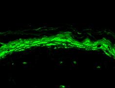 Immunohistochemistry analysis using Mouse Anti-Nav1.8 Monoclonal Antibody, Clone S134 (11523). Tissue: backskin. Species: Mouse. Fixation: Bouin’s Fixative and paraffin-embedded. Primary Antibody: Mouse Anti-Nav1.8 Monoclonal Antibody (11523) at 1:100 for 1 hour at RT. Secondary Antibody: FITC Goat Anti-Mouse (green) at 1:50 for 1 hour at RT. Localization: Heavy filaggrin-like staining, lower epidermal cells have some staining.
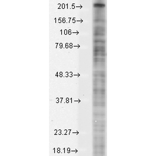 Western Blot analysis of Monkey COS transient cell lysate showing detection of Nav1.8 protein using Mouse Anti-Nav1.8 Monoclonal Antibody, Clone S134 (11523). Load: 15 µg. Block: 1.5% BSA for 30 minutes at RT. Primary Antibody: Mouse Anti-Nav1.8 Monoclonal Antibody (11523) at 1:1000 for 2 hours at RT. Secondary Antibody: Sheep Anti-Mouse IgG: HRP for 1 hour at RT.
 - -
- -
Antibody DetailsProduct DetailsReactive Species Human ⋅ Mouse ⋅ Rat Host Species Mouse Immunogen Fusion protein, aa 1724-1956 (cytoplasmic C-terminus) of rat Nav1.8 (accession no. Q62968). Product Concentration Lot Specific Formulation PBS, pH 7.4; 50% glycerol, 0.09% sodium azide. State of Matter Liquid Product Preparation Purified by Protein G affinity chromatography Storage and Handling This antibody is stable for at least one (1) year at -20°C. Country of Origin USA Shipping Next Day 2-8°C Applications and Recommended Usage? Quality Tested by Leinco Immunoblotting: use at 1-10ug/mL. A band of ~220kDa is detected.Immunohistochemistry and
Immunocytochemistry: use at 0.1-1ug/mL Immunofluorescence: use at 1-10ug/mL These are recommended concentrations. User should determine optimal concentrations for their application. Positive control: Rat dorsal root ganglia or lysate of COS cells transiently expressing Nav1.8. Each investigator should determine their own optimal working dilution for specific applications. See directions on lot specific datasheets, as information may periodically change. DescriptionDescriptionSpecificity Mouse Monoclonal Antibody specific to Nav1.8 Na+ Channel Background Ion channels are integral membrane proteins that help establish and control the small voltage gradient across the plasma membrane of living cells by allowing the flow of ions down their electrochemical gradient. Nav1.8 is a voltage-gated sodium channel that plays a critical role in the generation and conduction of action potentials and is thus important for electrical signaling by most excitable cells. Since Nav1.8 is not present in cardiac muscle or neurons in the CNS, blockers of Nav1.8 will not directly affect those cells and can have less side effects than current pain medications. Function Tetrodotoxin-resistant channel that mediates the voltage-dependent sodium ion permeability of excitable membranes. Assuming opened or closed conformations in response to the voltage difference across the membrane, the protein forms a sodium-selective channel through which sodium ions may pass in accordance with their electrochemical gradient. Plays a role in neuropathic pain mechanisms. {PubMed:12514212, PubMed:8538791, PubMed:8626372}. NCBI Gene Bank ID UniProt.org Research Area Ion Channels References & CitationsTechnical ProtocolsICC IF   AM Certificate of Analysis |
