

Anti-Notch1 Antibody (56544)


Anti-Notch1 Antibody (56544)
Product No.: 56544
- -
- -
Clone S253-32 Target Notch1 Formats AvailableView All Product Type Monoclonal Alternate Names Notch 1, Motch A, mT14, p300 [Cleaved into: Notch 1 extracellular truncation Isotype Mouse IgG1 Applications ICC , IF , WB |
Data
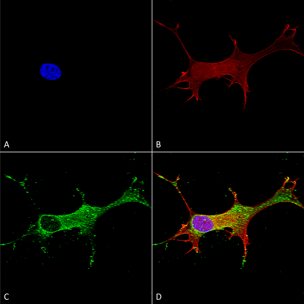 Immunocytochemistry/Immunofluorescence analysis using Mouse Anti-Notch1 Monoclonal Antibody, Clone S253-32 (56544). Tissue: Neuroblastoma cells (SH-SY5Y). Species: Human. Fixation: 4% PFA for 15 min. Primary Antibody: Mouse Anti-Notch1 Monoclonal Antibody (56544) at 1:50 for overnight at 4°C with slow rocking. Secondary Antibody: AlexaFluor 488 at 1:1000 for 1 hour at RT. Counterstain: Phalloidin-iFluor 647 (red) F-Actin stain; Hoechst (blue) nuclear stain at 1:800, 1.6mM for 20 min at RT. (A) Hoechst (blue) nuclear stain. (B) Phalloidin-iFluor 647 (red) F-Actin stain. (C) Notch1 Antibody (D) Composite.
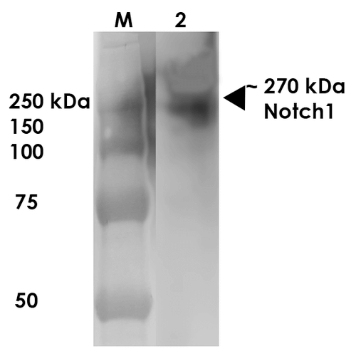 Western Blot analysis of Rat Brain Membrane showing detection of ~270 kDa Notch1 protein using Mouse Anti-Notch1 Monoclonal Antibody, Clone S253-32 (56544). Lane 1: MW Ladder. Lane 2: Rat Brain Membrane (10 µg). . Load: 10 µg. Block: 5% milk. Primary Antibody: Mouse Anti-Notch1 Monoclonal Antibody (56544) at 1:1000 for 1 hour at RT. Secondary Antibody: Goat Anti-Mouse IgG: HRP at 1:200 for 1 hour at RT. Color Development: TMB solution for 10 min at RT. Predicted/Observed Size: ~270 kDa.
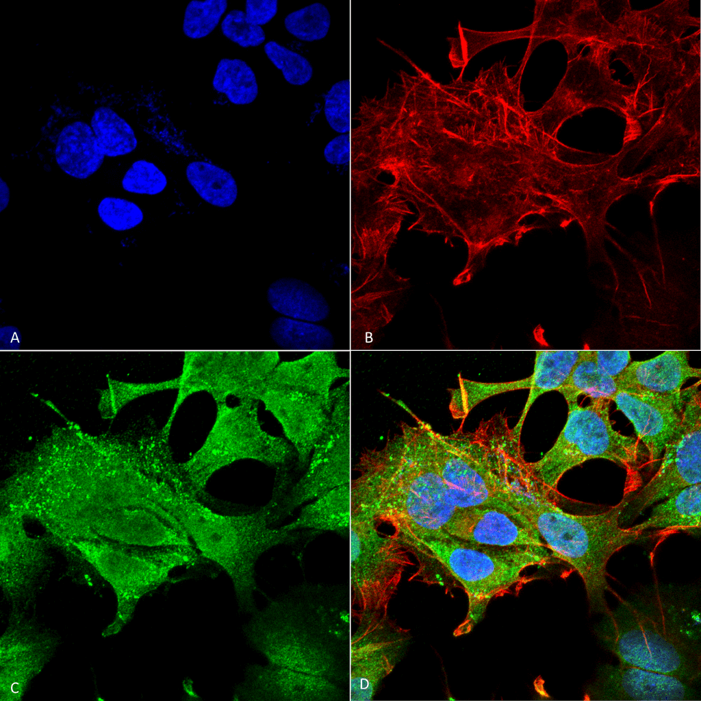 Immunocytochemistry/Immunofluorescence analysis using Mouse Anti-Notch1 Monoclonal Antibody, Clone S253-32 (56544). Tissue: Neuroblastoma cell line (SK-N-BE). Species: Human. Fixation: 4% Formaldehyde for 15 min at RT. Primary Antibody: Mouse Anti-Notch1 Monoclonal Antibody (56544) at 1:100 for 60 min at RT. Secondary Antibody: Goat Anti-Mouse ATTO 488 at 1:100 for 60 min at RT. Counterstain: Phalloidin Texas Red F-Actin stain; DAPI (blue) nuclear stain at 1:1000, 1:5000 for 60min RT, 5min RT. Localization: Cell Membrane, Nucleus. Magnification: 60X. (A) DAPI (blue) nuclear stain. (B) Phalloidin Texas Red F-Actin stain. (C) Notch1 Antibody. (D) Composite.
 - -
- -
Antibody DetailsProduct DetailsReactive Species Mouse ⋅ Rat Host Species Mouse Immunogen Fusion protein corresponding to aa 20-216 (extracellular N-terminus, EGF- like domains 1-5) of mouse Notch1 (accession no. Q01705). Product Concentration Lot Specific Formulation PBS, pH 7.4; 50% glycerol, 0.09% sodium azide. State of Matter Liquid Product Preparation Purified by Protein G affinity chromatography Storage and Handling This antibody is stable for at least one (1) year at -20°C. Avoid repeated freezing
and thawing. Regulatory Status For in vitro investigational use only. Not for
use in therapeutic or diagnostic procedures. Country of Origin USA Shipping Next Day 2-8°C Applications and Recommended Usage? Quality Tested by Leinco Immunoblotting: use at 1ug/mL. Bands of >270kDa, ~120kDa, and smaller fragments (due to proteolysis) are detected .
Positive control: Rat brain lysate. These are recommended concentrations. User should determine optimal concentrations for their application. Each investigator should determine their own optimal working dilution for specific applications. See directions on lot specific datasheets, as information may periodically change. DescriptionDescriptionSpecificity This antibody recognizes mouse and rat
Notch1. It does not cross-react with
Notch2 or Notch3. Background Members of the Notch trans-membrane protein family share structural characteristics including an extracellular domain consisting of multiple epidermal growth factor-like (EGF) repeats, and an intracellular domain consisting of multiple, different domain types. The Notch signaling network is an intercellular signaling pathway that regulates interactions between physically adjacent cells. The Notch1 protein is cleaved in the trans-Golgi network, and presented on the cell surface as a heterodimer. It functions as a receptor for membrane bound ligands, and may play multiple roles during development. There is evidence that activated Notch 1 and Notch 3 promote differentiation of progenitor cells into astroglia. Notch 1, activated before birth, induces radial glia differentiation,but postnatally induces the differentiation into astrocytes. Function Functions as a receptor for membrane-bound ligands Jagged-1 (JAG1), Jagged-2 (JAG2) and Delta-1 (DLL1) to regulate cell-fate determination. Upon ligand activation through the released notch intracellular domain (NICD) it forms a transcriptional activator complex with RBPJ/RBPSUH and activates genes of the enhancer of split locus. Affects the implementation of differentiation, proliferation and apoptotic programs. Involved in angiogenesis; negatively regulates endothelial cell proliferation and migration and angiogenic sprouting. Involved in the maturation of both CD4(+) and CD8(+) cells in the thymus. Important for follicular differentiation and possibly cell fate selection within the follicle. During cerebellar development, functions as a receptor for neuronal DNER and is involved in the differentiation of Bergmann glia. Represses neuronal and myogenic differentiation. May play an essential role in postimplantation development, probably in some aspect of cell specification and/or differentiation. May be involved in mesoderm development, somite formation and neurogenesis. May enhance HIF1A function by sequestering HIF1AN away from HIF1A. Required for the THBS4 function in regulating protective astrogenesis from the subventricular zone (SVZ) niche after injury. Involved in determination of left/right symmetry by modulating the balance between motile and immotile (sensory) cilia at the left-right organiser (LRO). {PubMed:15965470, PubMed:18299578, PubMed:23160044, PubMed:23615612}. NCBI Gene Bank ID UniProt.org Research Area Neuroscience References & CitationsTechnical ProtocolsICC IF  Certificate of Analysis |
Formats Available
 Products are for research use only. Not for use in diagnostic or therapeutic procedures.