

Anti-SUR2A [Clone S319A-14]


Anti-SUR2A [Clone S319A-14]
Product No.: 11570
- -
- -
Clone S319A-14 Target SUR2A Formats AvailableView All Product Type Monoclonal Alternate Names Sulfonylurea receptor 2 Isotype Mouse IgG2a Applications ICC , IF , IHC , WB |
Data
 Immunocytochemistry/Immunofluorescence analysis using Mouse Anti-SUR2A Monoclonal Antibody, Clone S319A-14 (11570). Tissue: Neuroblastoma cells (SH-SY5Y). Species: Human. Fixation: 4% PFA for 15 min. Primary Antibody: Mouse Anti-SUR2A Monoclonal Antibody (11570) at 1:200 for overnight at 4°C with slow rocking. Secondary Antibody: AlexaFluor 488 at 1:1000 for 1 hour at RT. Counterstain: Phalloidin-iFluor 647 (red) F-Actin stain; Hoechst (blue) nuclear stain at 1:800, 1.6mM for 20 min at RT. (A) Hoechst (blue) nuclear stain. (B) Phalloidin-iFluor 647 (red) F-Actin stain. (C) SUR2A Antibody (D) Composite.
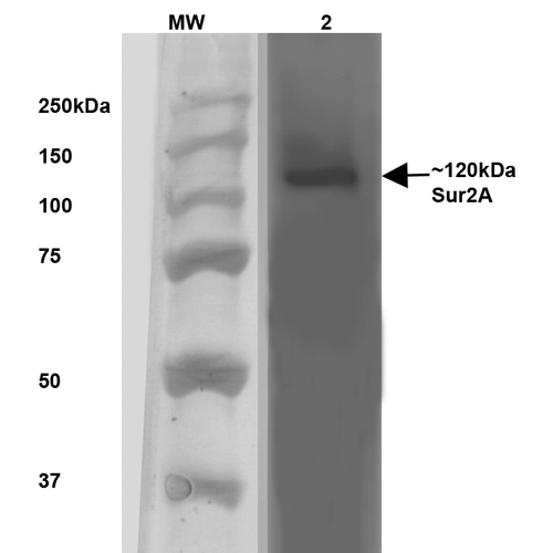 Western Blot analysis of Rat Brain Membrane showing detection of ~120 kDa SUR2A protein using Mouse Anti-SUR2A Monoclonal Antibody, Clone S319A-14 (11570). Lane 1: MW Ladder. Lane 2: Rat Brain Membrane (10 µg). . Load: 10 µg. Block: 5% milk. Primary Antibody: Mouse Anti-SUR2A Monoclonal Antibody (11570) at 1:1000 for 1 hour at RT. Secondary Antibody: Goat Anti-Mouse IgG: HRP at 1:200 for 1 hour at RT. Color Development: TMB solution for 10 min at RT. Predicted/Observed Size: ~120 kDa.
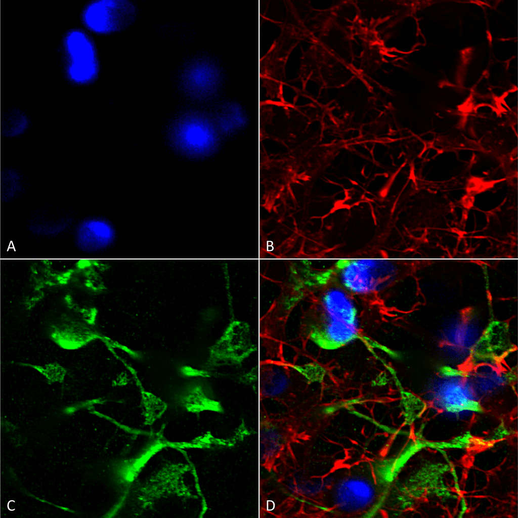
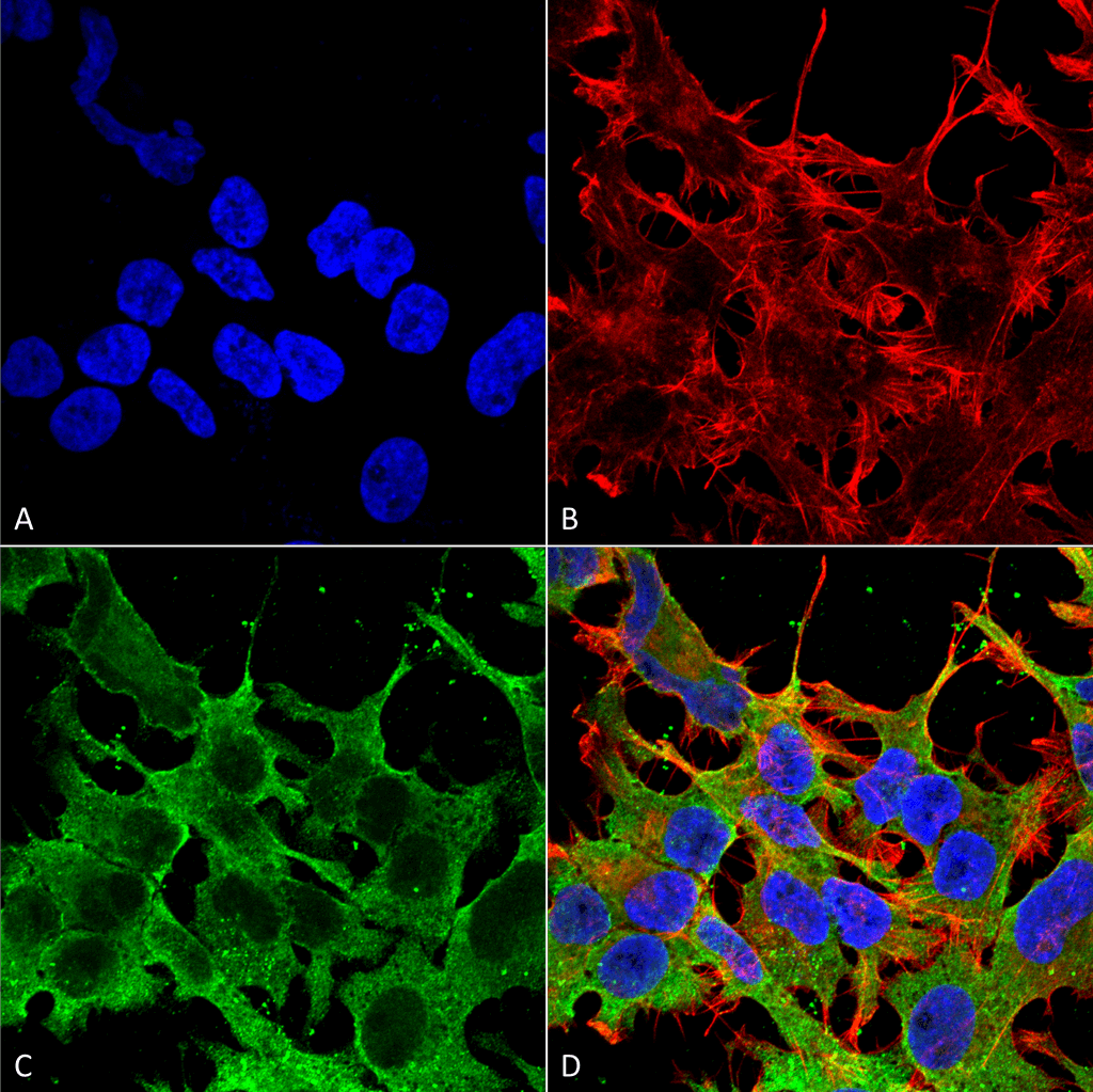 Immunocytochemistry/Immunofluorescence analysis using Mouse Anti-SUR2A Monoclonal Antibody, Clone S319A-14 (11570). Tissue: Neuroblastoma cell line (SK-N-BE). Species: Human. Fixation: 4% Formaldehyde for 15 min at RT. Primary Antibody: Mouse Anti-SUR2A Monoclonal Antibody (11570) at 1:100 for 60 min at RT. Secondary Antibody: Goat Anti-Mouse ATTO 488 at 1:100 for 60 min at RT. Counterstain: Phalloidin Texas Red F-Actin stain; DAPI (blue) nuclear stain at 1:1000, 1:5000 for 60min RT, 5min RT. Localization: Membrane. Magnification: 60X. (A) DAPI (blue) nuclear stain. (B) Phalloidin Texas Red F-Actin stain. (C) SUR2A Antibody. (D) Composite.
 - -
- -
Antibody DetailsProduct DetailsReactivity Species Mouse ⋅ Rat Host Species Mouse Immunogen Fusion protein corresponding to aa 1505-1546 (SSIVDAGLVLVFSEGILVECDTGPNLLQ HKNGLFSTLVMTNK, cytoplasmic C- terminus) of mouse SUR2A (accession no. P70170). Product Concentration Lot Specific Formulation PBS, pH 7.4; 50% glycerol, 0.09% sodium azide. State of Matter Liquid Product Preparation Purified by Protein G affinity chromatography Storage and Handling This antibody is stable for at least one (1) year at -20°C. Avoid repeated freeze-thaw cycles. Regulatory Status For in vitro investigational use only. Not for use in therapeutic or diagnostic procedures. Country of Origin USA Shipping Next Day 2-8°C Applications and Recommended Usage? Quality Tested by Leinco Immunoblotting: use at 1-2ug/mL. A band of ~120kDa is detected.
Positive control: Mouse brain lysate. These are recommended concentrations. User should determine optimal concentrations for their application. Each investigator should determine their own optimal working dilution for specific applications. See directions on lot specific datasheets, as information may periodically change. DescriptionSpecificity This antibody recognizes mouse and rat SUR2A. It does not cross-react with SUR2B. Background Sulfonylurea receptors (SUR) are membrane proteins that are molecular targets of the sulfonylurea class of antidiabetic drugs whose mechanism of action is to promote insulin release from pancreatic beta cells. SUR proteins are subunits of the inward-rectifier potassium ion channels Kir6.x (6.1 and 6.2). The association of four Kir6.x and four SUR subunits form an ion conducting channel commonly referred to as the KATP channel. The primary function of the SUR is to sense intracellular levels of ATP and ADP and facilitate the opening or closing of its associated Kir6.x potassium channel, thus monitoring the energy balance within cells. Antigen DetailsFunction Subunit of ATP-sensitive potassium channels (KATP). Can form cardiac and smooth muscle-type KATP channels with KCNJ11. KCNJ11 forms the channel pore while ABCC9 is required for activation and regulation. NCBI Gene Bank ID UniProt.org Research Area Ion Channels References & CitationsTechnical ProtocolsICC IF   |
Formats Available
 Products are for research use only. Not for use in diagnostic or therapeutic procedures.