

Anti-TrpC7 Ca+2 Channel [Clone S64A-36]


Anti-TrpC7 Ca+2 Channel [Clone S64A-36]
Product No.: 11522
- -
- -
Clone S64A-36 Target TrpC7 Ca+2 Channel Formats AvailableView All Product Type Monoclonal Alternate Names TrpC7, Transient receptor protein 7, TRP-7, hTRP7 Isotype Mouse IgG1 Applications ICC , IF , IHC , IP , WB , AM |
Data
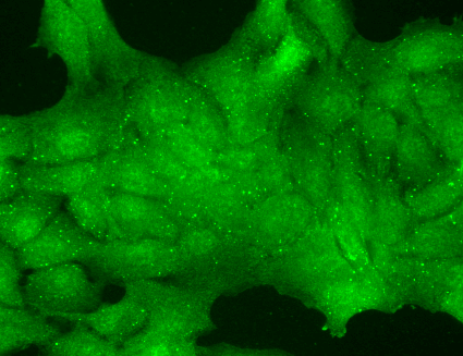 Immunocytochemistry/Immunofluorescence analysis using Mouse Anti-TrpC7 Monoclonal Antibody, Clone N64A/36 (11522). Tissue: HaCaT cells. Species: Human. Fixation: Cold 100% methanol for 10 minutes at -20°C. Primary Antibody: Mouse Anti-TrpC7 Monoclonal Antibody (11522) at 1:100 for 1 hour at RT. Secondary Antibody: FITC Goat Anti-Mouse (green) at 1:50 for 1 hour at RT. Localization: Nuclear staining .
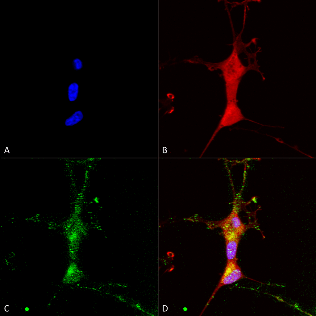 Immunocytochemistry/Immunofluorescence analysis using Mouse Anti-TrpC7 Monoclonal Antibody, Clone N64A/36 (11522). Tissue: Neuroblastoma cells (SH-SY5Y). Species: Human. Fixation: 4% PFA for 15 min. Primary Antibody: Mouse Anti-TrpC7 Monoclonal Antibody (11522) at 1:100 for overnight at 4°C with slow rocking. Secondary Antibody: AlexaFluor 488 at 1:1000 for 1 hour at RT. Counterstain: Phalloidin-iFluor 647 (red) F-Actin stain; Hoechst (blue) nuclear stain at 1:800, 1.6mM for 20 min at RT. (A) Hoechst (blue) nuclear stain. (B) Phalloidin-iFluor 647 (red) F-Actin stain. (C) TrpC7 Antibody (D) Composite.
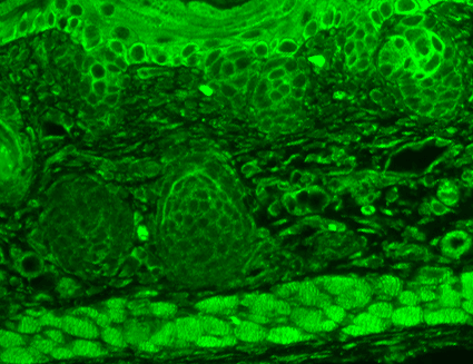 Immunohistochemistry analysis using Mouse Anti-TrpC7 Monoclonal Antibody, Clone N64A/36 (11522). Tissue: backskin. Species: Mouse. Fixation: Bouin’s Fixative and paraffin-embedded. Primary Antibody: Mouse Anti-TrpC7 Monoclonal Antibody (11522) at 1:100 for 1 hour at RT. Secondary Antibody: FITC Goat Anti-Mouse (green) at 1:50 for 1 hour at RT. Localization: Everything.
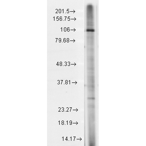 Western Blot analysis of Rat brain membrane lysate showing detection of TrpC7 protein using Mouse Anti-TrpC7 Monoclonal Antibody, Clone N64A/36 (11522). Load: 15 µg. Block: 1.5% BSA for 30 minutes at RT. Primary Antibody: Mouse Anti-TrpC7 Monoclonal Antibody (11522) at 1:1000 for 2 hours at RT. Secondary Antibody: Sheep Anti-Mouse IgG: HRP for 1 hour at RT.
 - -
- -
Antibody DetailsProduct DetailsReactivity Species Human ⋅ Mouse ⋅ Rat Host Species Mouse Immunogen Synthetic peptide corresponding to aa 845-862 (FGNLNKDHLRVNKGKDI) of human TrpC7 (accession no. Q9HCX4). This sequence is 100 Product Concentration Lot Specific Formulation PBS, pH 7.4; 50% glycerol, 0.09% sodium azide. State of Matter Liquid Product Preparation Purified by Protein G affinity chromatography Storage and Handling This antibody is stable for at least one (1) year at -20°C. Avoid repeated freeze-thaw cycles.
Regulatory Status For in vitro investigational use only. Not for use in therapeutic or diagnostic procedures. Country of Origin USA Shipping Next Day 2-8°C Applications and Recommended Usage? Quality Tested by Leinco Immunoblotting: use at 1-10ug/mL. A band of ~100kDa is detected.Immunohistochemistry and
Immunocytochemistry: use at 0.1-1ug/mL Immunofluorescence: use at 1-10ug/mL These are recommended concentrations. User should determine optimal concentrations for their application. Positive control: Rat brain lysate Each investigator should determine their own optimal working dilution for specific applications. See directions on lot specific datasheets, as information may periodically change. DescriptionSpecificity This antibody recognizes human, mouse, and rat TrpC7. Background Ion channels are integral membrane proteins that help establish and control the small voltage gradient across the plasma membrane of living cells by allowing the flow of ions down their electrochemical gradient. Transient receptor potential cation channel, subfamily C, member 7 (TrpC7) has non-selective cation constitutive activity and susceptibility to negative regulation by extracellular Ca+2. TrpC7 plays an important role in the Ca+2 signaling pathway. TrpC7 is abundantly expressed in heart and may contribute to heart failure. Antigen DetailsFunction Thought to form a receptor-activated non-selective calcium permeant cation channel. Probably is operated by a phosphatidylinositol second messenger system activated by receptor tyrosine kinases or G-protein coupled receptors. Activated by diacylglycerol (DAG) (By similarity). May also be activated by intracellular calcium store depletion. {ECO:0000250}. NCBI Gene Bank ID UniProt.org Research Area Ion Channels References & CitationsTechnical ProtocolsICC IF    AM |
Formats Available
 Products are for research use only. Not for use in diagnostic or therapeutic procedures.