Epidermal growth factor (EGF), also known as Urogastrone, is a growth factor that plays an important role in the regulation of cell growth, proliferation and differentiation (1). It is produced by many cell types, in blood and various body fluids, including milk, urine, saliva, seminal fluid, pancreatic juice, cerebrospinal fluid, and amniotic fluid (1). EGF acts by binding with high affinity to epidermal growth factor receptor (EGFR) on the cell surface, activating an extensive network of signal transduction pathways that include PI3K/AKT, RAS/ERK and JAK/STAT. It is also a mitogen for fibroblasts, epithelial and endothelial cells, and promotes colony formation of epiderma. EGF has been shown to regulate tumor cell invasion through MMP-2 activation in various tumor cell types (2). Additionally, EGF has been shown to inhibit gastric secretion, and to be involved in wound healing (3). Because of its key role in driving the proliferation of cells, EGFR is a target of several anti-cancer drugs currently in development.
Protein Details
Purity
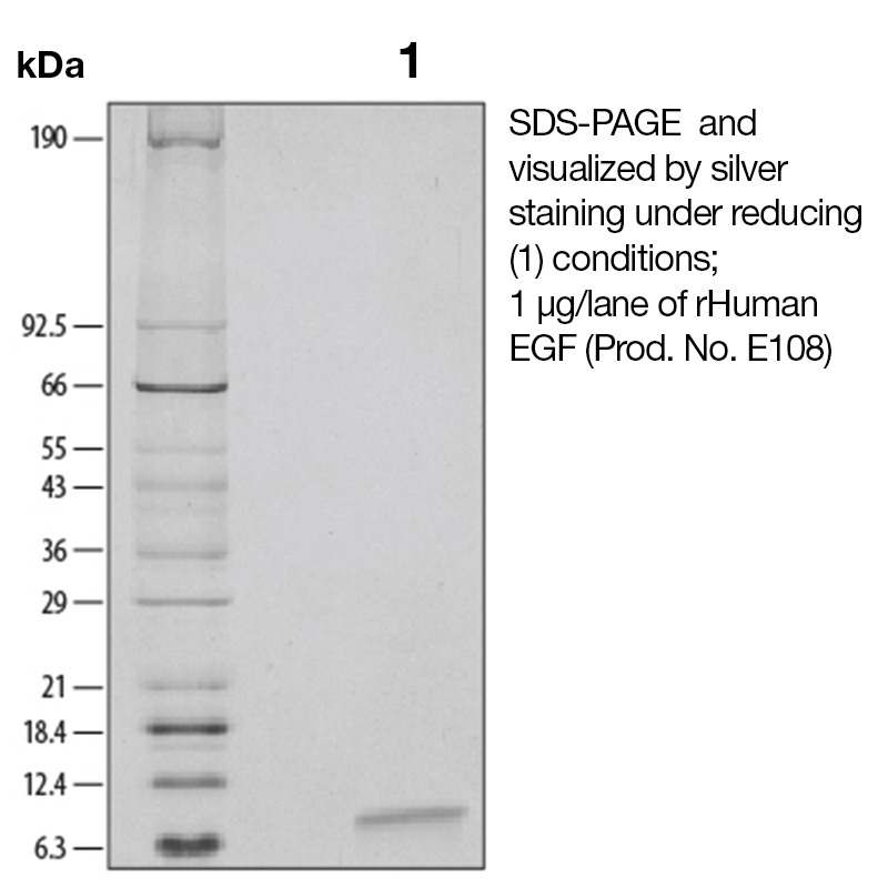
>97% by SDS-PAGE and analyzed by silver stain.
Endotoxin Level
<0.1 EU/µg as determined by the LAL method
Biological Activity
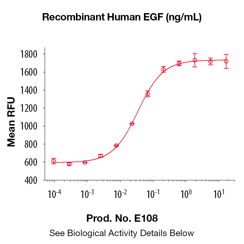
The biological activity of Human EGF was determined by its ability to stimulate proliferation in an EGF-responsive mouse fibroblast cell line, Balb/3T3 (Rubin, J.S. et al., 1991, Proc. Natl. Acad. Sci. USA 88:415). The expected ED<sub>50</sub> is typically 20 - 100 pg/ml.
The predicted molecular weight of Recombinant Human EGF is Mr 6 kDa. Additionally, the actual molecular weight as observed by migration on SDS-PAGE is 6 kDa (reducing conditions).
Predicted Molecular Mass
6
Formulation
This recombinant protein was 0.2 µm filtered and lyophilized from modified Dulbecco’s phosphate buffered saline (1X PBS) pH 7.2 – 7.3 with no calcium, magnesium, or preservatives.
Storage and Stability
This lyophilized protein is stable for six to twelve months when stored desiccated at -20°C to -70°C. After aseptic reconstitution, this protein may be stored at 2°C to 8°C for one month or at -20°C to -70°C in a manual defrost freezer. Avoid Repeated Freeze Thaw Cycles. See Product Insert for exact lot specific storage instructions.
Powered by AI: AI is experimental and still learning how to provide the best assistance. It may occasionally generate incorrect or incomplete responses. Please do not rely solely on its recommendations when making purchasing decisions or designing experiments.
Recombinant Human EGF (Epidermal Growth Factor) is widely used in research because it is a potent, well-characterized mitogen that promotes cell proliferation, migration, and differentiation, making it essential for studies in cell biology, tissue engineering, wound healing, and organoid culture.
Key reasons to use recombinant human EGF in research applications:
Promotes Cell Proliferation and Viability: EGF binds to the EGF receptor (EGFR), activating downstream signaling pathways (such as ERK1/2 and c-Jun), which drive cell proliferation, survival, and differentiation in various cell types, including epithelial cells, fibroblasts, and stem cells.
Enhances Cell Migration and Wound Healing: EGF stimulates cell migration and extracellular matrix synthesis, accelerating wound closure and tissue regeneration in both in vitro and in vivo models.
Supports Organoid and 3D Cell Culture: Recombinant EGF is critical for the growth and maintenance of organoids and 3D tissue models, providing high bioactivity and batch-to-batch consistency, which are essential for reproducible results in advanced cell culture systems.
Enables Controlled Experimental Conditions: Recombinant production ensures high purity, defined activity, and minimal contamination, reducing variability compared to native or animal-derived growth factors.
Facilitates Mechanistic Studies: EGF is used to dissect signaling pathways involved in cell growth, differentiation, and disease processes, including cancer biology, tissue regeneration, and developmental biology.
Versatility Across Applications: EGF is used in assays for cell proliferation, migration (e.g., scratch assays), colony formation, and as a supplement in serum-free media for stem cell and primary cell culture.
Clinical and Translational Relevance: EGF’s roles in wound healing, tissue repair, and as a potential therapeutic agent make it valuable for translational research and preclinical studies.
In summary, recombinant human EGF is a reliable, potent, and versatile tool for stimulating cell growth and studying cellular processes in a wide range of research applications.
Yes, recombinant human EGF can be used as a standard for quantification or calibration in ELISA assays, provided it is of high purity and properly validated for this application.
Recombinant human EGF is commonly employed as a standard in ELISA protocols to generate a calibration curve for quantifying EGF concentrations in biological samples. The protein should be highly pure (typically >98% by SDS-PAGE and HPLC), and its concentration must be accurately determined after reconstitution. ELISA kits designed for EGF quantification routinely use recombinant human EGF as the standard, and validation studies show that the assay can reliably measure both recombinant and natural EGF, with parallel linearity and recovery rates between 89–111%.
Key considerations for use:
Ensure the recombinant EGF is reconstituted and diluted according to the ELISA kit instructions, using the recommended calibrator diluent for your sample type.
The standard curve should cover the expected range of EGF concentrations in your samples, typically from low pg/mL to ng/mL levels.
The recombinant EGF standard should be stored and handled as specified to maintain stability and activity.
For research use only; not for diagnostic procedures unless specifically validated.
Best practices:
Use the same source and lot of recombinant EGF for all calibration curves within a study to ensure consistency.
Confirm that the recombinant EGF standard is calibrated against an international reference, such as the WHO International Standard (NIBSC code: 91/530), for highest accuracy.
Validate parallelism between the standard curve and sample dilution curves to confirm assay reliability.
In summary, recombinant human EGF is suitable and widely accepted as a standard for ELISA quantification, provided it meets purity, calibration, and validation requirements for your specific assay system.
Recombinant Human EGF (rhEGF) has been validated in published research for a range of applications, primarily focused on cell biology, tissue engineering, and regenerative medicine. Key validated applications include:
Cell proliferation and viability assays: rhEGF has been shown to significantly promote the proliferation and viability of various cell types, including fibroblasts (e.g., NIH 3T3) and epithelial cells (e.g., HaCaT), by activating intracellular signaling pathways such as ERK1/2 and c-Jun.
Wound healing and tissue regeneration models: rhEGF is widely used in both in vitro and in vivo models to accelerate wound closure, enhance tissue repair, and minimize scar formation. Its effects have been validated in studies of skin wound healing, burns, and chronic ulcers.
Gene expression and signaling pathway analysis: RNA sequencing and protein expression studies have validated that rhEGF upregulates genes and proteins involved in cell growth, DNA replication, ribosome biogenesis, and cell cycle progression.
Stem cell culture and differentiation: rhEGF is used to stimulate proliferation and differentiation of stem cells, particularly in neural and intestinal stem cell cultures.
Functional assays and biomaterial studies: rhEGF has been incorporated into biomaterials for controlled release in tissue engineering applications, and its biological activity has been validated in these contexts.
Immunoassays and biochemical assays: rhEGF is used as a standard or stimulant in ELISA, Western blot, and immunofluorescence assays to study EGF receptor signaling and downstream effects.
Additional validated uses include:
Cosmetic and dermatological research: rhEGF is studied for its effects on skin rejuvenation, anti-aging, and cosmetic wound healing.
Disease modeling: rhEGF is used to investigate EGF signaling in cancer, renal disease, and developmental biology.
Summary Table of Validated Applications
Application Area
Example Assays/Models
References
Cell proliferation/viability
MTT, cell counting, signaling activation
Wound healing/tissue regeneration
In vitro scratch, animal wound models
Gene/protein expression
RNA-seq, qPCR, Western blot, immunofluorescence
Stem cell culture/differentiation
Neural/intestinal stem cell expansion
Functional/biomaterial assays
Controlled release, tissue engineering
Immunoassays
ELISA, Western blot, immunofluorescence
Cosmetic/dermatological research
Skin models, cosmetic wound healing
Disease modeling
Cancer, renal, developmental studies
These applications are supported by both direct experimental validation and widespread use in published research.
To reconstitute and prepare Recombinant Human EGF (Epidermal Growth Factor) protein for cell culture experiments, follow these best practices based on manufacturer recommendations and scientific protocols:
1. Reconstitution
Centrifuge the vial briefly before opening to ensure all lyophilized powder is at the bottom.
Reconstitute the lyophilized EGF in sterile, endotoxin-free water, PBS, or an appropriate aqueous buffer (e.g., 10 mM acetic acid or PBS containing 0.1% BSA).
Gently mix after reconstitution; avoid vigorous shaking to prevent protein denaturation.
2. Preparation for Cell Culture
Aliquot the reconstituted EGF into small working volumes to minimize freeze-thaw cycles.
Store aliquots at ≤ –20°C for long-term storage. Avoid frost-free freezers.
For short-term use (within 1 month), store at 4°C.
Avoid repeated freeze-thaw cycles, as this can reduce bioactivity.
3. Dilution for Cell Culture
Dilute the reconstituted EGF in low endotoxin medium or a buffered solution (e.g., PBS or culture medium) containing a carrier protein such as heat-inactivated fetal calf serum (FCS) or tissue culture-grade BSA (0.1%).
Typical working concentrations for cell culture range from 0.5–25 ng/mL, but the optimal concentration should be determined for your specific cell type and application using a dose-response assay.
4. Additional Tips
Microcentrifuge the solution before use if a precipitate is observed.
Sterility: Ensure all solutions and equipment are sterile to prevent contamination in cell culture.
Endotoxin: Use low endotoxin reagents to minimize unwanted immune responses in sensitive cultures.
Summary Protocol:
Centrifuge vial briefly.
Reconstitute in sterile water or PBS (0.1–1.0 mg/mL).
Gently mix; aliquot and store at ≤ –20°C.
Dilute in culture medium or buffer with 0.1% BSA for use.
Use freshly thawed aliquots; avoid repeated freeze-thaw cycles.
These steps will help maintain the bioactivity and stability of recombinant human EGF for reliable results in cell culture experiments.
References & Citations
1. Carpenter, G. et al. (1986) J. Biol. Chem. 265:770
2. Cosen-Binker, LI. et al. (2009) Biochem. Biophys. Res. Commun. 379:445
3. Machowska, A. et al. (2008) Inflammopharmacol. 16:40


 Products are for research use only. Not for use in diagnostic or therapeutic procedures.