

Anti-Hsp25/Hsp27 [Clone 8A7]

Anti-Hsp25/Hsp27 [Clone 8A7]
Product No.: 11110
- -
- -
Clone 8A7 Target Hsp25/Hsp27 Formats AvailableView All Product Type Monoclonal Alternate Names HspB1, 28 kDa heat shock protein, Estrogen-regulated 24 kDa protein, Heat shock 27 kDa protein, HSP 27, Stress-responsive protein 27, SRP27 Isotype Mouse IgG1 Applications FACS , ICC , IF , IHC , IP , WB , FCM |
Data
 Immunohistochemistry analysis using Mouse Anti-Hsp27 Monoclonal Antibody, Clone 8A7 (11110). Tissue: colon carcinoma. Species: Human. Fixation: Formalin. Primary Antibody: Mouse Anti-Hsp27 Monoclonal Antibody (11110) at 1:5000 for 12 hours at 4°C. Secondary Antibody: Biotin Goat Anti-Mouse at 1:2000 for 1 hour at RT. Counterstain: Mayer Hematoxylin (purple/blue) nuclear stain at 200 µl for 2 minutes at RT. Localization: Inflammatory cells. Magnification: 40x.
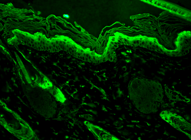 Immunohistochemistry analysis using Mouse Anti-Hsp27 Monoclonal Antibody, Clone 8A7 (11110). Tissue: backskin. Species: Mouse. Fixation: Bouin’s Fixative and paraffin-embedded. Primary Antibody: Mouse Anti-Hsp27 Monoclonal Antibody (11110) at 1:100 for 1 hour at RT. Secondary Antibody: FITC Goat Anti-Mouse (green) at 1:50 for 1 hour at RT. Localization: Epidermis.
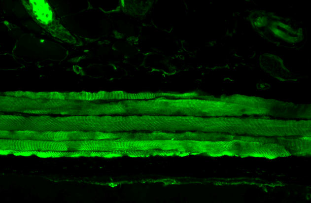 Immunohistochemistry analysis using Mouse Anti-Hsp27 Monoclonal Antibody, Clone 8A7 (11110). Tissue: backskin. Species: Mouse. Fixation: Bouin’s Fixative and paraffin-embedded. Primary Antibody: Mouse Anti-Hsp27 Monoclonal Antibody (11110) at 1:100 for 1 hour at RT. Secondary Antibody: FITC Goat Anti-Mouse (green) at 1:50 for 1 hour at RT. Localization: Epidermis.
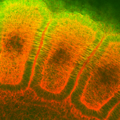 Immunohistochemistry analysis using Mouse Anti-Hsp27 Monoclonal Antibody, Clone 8A7 (11110). Tissue: embryo somites. Species: Rat. Primary Antibody: Mouse Anti-Hsp27 Monoclonal Antibody (11110) at 1:1000. Secondary Antibody: FITC Goat Anti-Mouse (green). Counterstain: Rhodamine-phalloidin labeled actin (red). Investigation at 11 days gestation. Courtesy of: Mike Welsh, Umich.
 - -
- -
Antibody DetailsProduct DetailsReactivity Species Bovine ⋅ Canine ⋅ Guinea Pig ⋅ Hamster ⋅ Human ⋅ Mouse ⋅ Rat Host Species Mouse Immunogen Hsp27 peptide Product Concentration Lot Specific Formulation PBS, pH 7.4. State of Matter Liquid Product Preparation Purified by Protein G affinity chromatography Storage and Handling This antibody is stable for at least one (1) year at -20°C. Regulatory Status For in vitro investigational use only. Not for
use in therapeutic or diagnostic procedures. Country of Origin USA Shipping Next Day 2-8°C Applications and Recommended Usage? Quality Tested by Leinco Immunoblotting: use at 0.2ug/mL (ECL). A band of 25-27 kDa is detected.
Immunofluorescence: use at 5ug/mL. Positive control: HeLa cell lysate Each investigator should determine their own optimal working dilution for specific applications. See directions on lot specific datasheets, as information may periodically change. DescriptionSpecificity This antibody recognizes human,
mouse, rat, bovine, canine, guinea pig,
and hamster Hsp 25/27. Some crossreactivity with alpha B crystallin. Background Hsp25 is the mouse homologue of human Hsp27, a member of the small heat shock protein family comprised of proteins of ~15->30kDa. Oligomers of Hsp27 consist of as many as 8-40 Hsp27 monomers; large oligomers have high chaperone activity whereas dimers have no chaperone activity. Hsp27 is localized to the cytoplasm of unstressed cells but redistributes to the nucleus in response to stress where it may stabilize DNA and/or the nuclear membrane. It can be rapidly phosphorylated in response to physiological stimuli, and, therefore, has been suggested as an important intermediate in second messenger- mediated signaling pathways. Hsp27 appears to be involved in cell differentiation and may play a role in termination of cell growth. Antigen DetailsFunction Small heat shock protein which functions as a molecular chaperone probably maintaining denatured proteins in a folding-competent state (PubMed:10383393, PubMed:20178975). Plays a role in stress resistance and actin organization (PubMed:19166925). Through its molecular chaperone activity may regulate numerous biological processes including the phosphorylation and the axonal transport of neurofilament proteins (PubMed:23728742). {PubMed:10383393, PubMed:19166925, PubMed:20178975, PubMed:23728742}. NCBI Gene Bank ID UniProt.org Research Area Heat Shock & Stress Proteins References & CitationsTechnical ProtocolsFACS ICC IF    FCM |
