

Anti-α 1 GABA-A Receptor [Clone S95-35]


Anti-α 1 GABA-A Receptor [Clone S95-35]
Product No.: 56467
Data
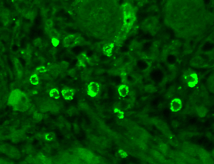 Immunohistochemistry analysis using Mouse Anti-GABA A Receptor Monoclonal Antibody, Clone N95/35 (56467). Tissue: backskin. Species: Mouse. Fixation: Bouin’s Fixative and paraffin-embedded. Primary Antibody: Mouse Anti-GABA A Receptor Monoclonal Antibody (56467) at 1:100 for 1 hour at RT. Secondary Antibody: FITC Goat Anti-Mouse (green) at 1:50 for 1 hour at RT. Localization: Dermal Cells.
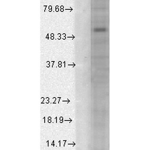 Western Blot analysis of Human Cell line lysates showing detection of GABA A Receptor protein using Mouse Anti-GABA A Receptor Monoclonal Antibody, Clone N95/35 (56467). Load: 15 µg. Block: 1.5% BSA for 30 minutes at RT. Primary Antibody: Mouse Anti-GABA A Receptor Monoclonal Antibody (56467) at 1:1000 for 2 hours at RT. Secondary Antibody: Sheep Anti-Mouse IgG: HRP for 1 hour at RT.
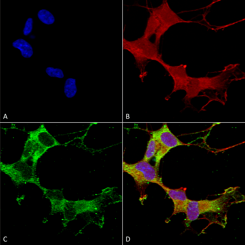 Immunocytochemistry/Immunofluorescence analysis using Mouse Anti-GABA-A Receptor Alpha-1 Monoclonal Antibody, Clone N95/35 (56467). Tissue: Neuroblastoma cells (SH-SY5Y). Species: Human. Fixation: 4% PFA for 15 min. Primary Antibody: Mouse Anti-GABA-A Receptor Alpha-1 Monoclonal Antibody (56467) at 1:50 for overnight at 4°C with slow rocking. Secondary Antibody: AlexaFluor 488 at 1:1000 for 1 hour at RT. Counterstain: Phalloidin-iFluor 647 (red) F-Actin stain; Hoechst (blue) nuclear stain at 1:800, 1.6mM for 20 min at RT. (A) Hoechst (blue) nuclear stain. (B) Phalloidin-iFluor 647 (red) F-Actin stain. (C) GABA-A Receptor Alpha-1 Antibody (D) Composite.
 - -
- -
Antibody DetailsProduct DetailsReactive Species Human ⋅ Mouse ⋅ Rat Host Species Mouse Immunogen Fusion protein corresponding to aa 355-394 of mouse Alpha 1 GABA-A receptor (accession no. P62812). Product Concentration Lot Specific Formulation PBS, pH 7.4; 50% glycerol, 0.09% sodium azide. Purified by Protein G affinity chromatography. State of Matter Liquid Product Preparation Purified by Protein G affinity chromatography Storage and Handling This antibody is stable for at least one (1) year at -20°C. Avoid repeated freezing
and thawing. Regulatory Status For in vitro investigational use only. Not for
use in therapeutic or diagnostic procedures. Country of Origin USA Shipping Next Day 2-8°C Applications and Recommended Usage? Quality Tested by Leinco Immunoblotting: use at 1-10ug/mL. A band of ~55kDa is detected.Immunohistochemistry and
Immunocytochemistry: use at 0.1-1ug/mL Immunofluorescence: use at 1-10ug/mL These are recommended concentrations. User should determine optimal concentrations for their application. Positive control: Rat brain lysate. Each investigator should determine their own optimal working dilution for specific applications. See directions on lot specific datasheets, as information may periodically change. DescriptionDescriptionSpecificity This antibody recognizes human, mouse,
and rat Alpha 1 GABA-A receptor. It does
not cross-react with Alpha 2 or Alpha 3
GABA-A receptors. Background GABA is the major inhibitory neuro- transmitter in the vertebrate brain. GABA receptors consist of five subunits that form a chloride ion channel. Subunit families alpha, beta, g, and delta have been studied extensively; π and epsilon have been identified recently. Studies of recombinant GABA receptors have shown that individual subunits and their subtypes confer different sensitivities to GABA receptor modulators. Subunit subtypes are differentially expressed throughout development and in different CNS regions, reducing the total number of possible isoforms that can be formed in different brain regions and in individual cells. Function Ligand-gated chloride channel which is a component of the heteropentameric receptor for GABA, the major inhibitory neurotransmitter in the brain (PubMed:23909897, PubMed:25489750, PubMed:29950725). Plays an important role in the formation of functional inhibitory GABAergic synapses in addition to mediating synaptic inhibition as a GABA-gated ion channel (PubMed:23909897, PubMed:25489750). The gamma2 subunit is necessary but not sufficient for a rapid formation of active synaptic contacts and the synaptogenic effect of this subunit is influenced by the type of alpha and beta subunits present in the receptor pentamer (By similarity). The alpha1/beta2/gamma2 receptor and the alpha1/beta3/gamma2 receptor exhibit synaptogenic activity (PubMed:23909897, PubMed:25489750). GABRA1-mediated plasticity in the orbitofrontal cortex regulates context-dependent action selection (By similarity). Functions also as histamine receptor and mediates cellular responses to histamine (By similarity). {UniProtKB:P62812, UniProtKB:P62813, PubMed:23909897, PubMed:25489750, PubMed:29950725}. NCBI Gene Bank ID UniProt.org Research Area Neuroscience References & CitationsTechnical ProtocolsICC IF   AM Certificate of Analysis |
Formats Available
 Products are for research use only. Not for use in diagnostic or therapeutic procedures.