

Anti-Ataxin-1 Antibody (56549)


Anti-Ataxin-1 Antibody (56549)
Product No.: 56549
- -
- -
Clone S65-37 Target Ataxin-1 Formats AvailableView All Product Type Monoclonal Alternate Names Spinocerebellar ataxia type 1 protein homolog Isotype Mouse IgG1 Applications ICC , IF , WB |
Data
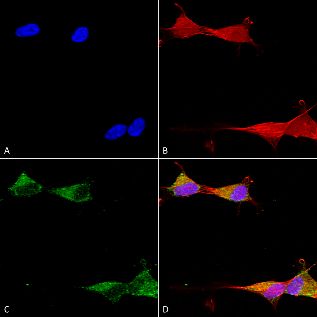 Immunocytochemistry/Immunofluorescence analysis using Mouse Anti-Ataxin 1 Monoclonal Antibody, Clone S65-37 (56549). Tissue: Neuroblastoma cells (SH-SY5Y). Species: Human. Fixation: 4% PFA for 15 min. Primary Antibody: Mouse Anti-Ataxin 1 Monoclonal Antibody (56549) at 1:50 for overnight at 4°C with slow rocking. Secondary Antibody: AlexaFluor 488 at 1:1000 for 1 hour at RT. Counterstain: Phalloidin-iFluor 647 (red) F-Actin stain; Hoechst (blue) nuclear stain at 1:800, 1.6mM for 20 min at RT. (A) Hoechst (blue) nuclear stain. (B) Phalloidin-iFluor 647 (red) F-Actin stain. (C) Ataxin 1 Antibody (D) Composite.
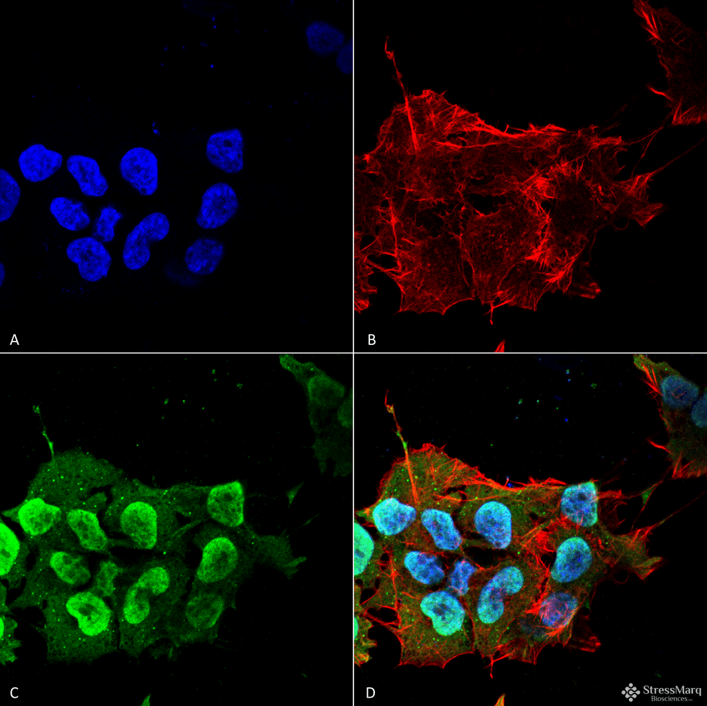 Immunocytochemistry/Immunofluorescence analysis using Mouse Anti-Ataxin 1 Monoclonal Antibody, Clone S65-37 (56549). Tissue: Neuroblastoma cell line (SK-N-BE). Species: Human. Fixation: 4% Formaldehyde for 15 min at RT. Primary Antibody: Mouse Anti-Ataxin 1 Monoclonal Antibody (56549) at 1:100 for 60 min at RT. Secondary Antibody: Goat Anti-Mouse ATTO 488 at 1:100 for 60 min at RT. Counterstain: Phalloidin Texas Red F-Actin stain; DAPI (blue) nuclear stain at 1:1000; 1:5000 for 60 min RT, 5 min RT. Localization: Cytoplasm, Nucleus. Magnification: 60X. (A) DAPI (blue) nuclear stain. (B) Phalloidin Texas Red F-Actin stain. (C) Ataxin 1 Antibody. (D) Composite.
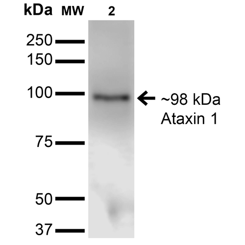 Western Blot analysis of Monkey COS-1 cells transfected with Ataxin- 1 showing detection of ~85 kDa Ataxin 1 protein using Mouse Anti-Ataxin 1 Monoclonal Antibody, Clone S65-37 (56549). Lane 1: Molecular Weight Ladder. Lane 2: Monkey COS-1 cells transfected with Ataxin- 1. Load: 15 µg. Block: 2% BSA and 2% Skim Milk in 1X TBST. Primary Antibody: Mouse Anti-Ataxin 1 Monoclonal Antibody (56549) at 1:200 for 16 hours at 4°C. Secondary Antibody: Goat Anti-Mouse IgG: HRP at 1:1000 for 1 hour RT. Color Development: ECL solution for 6 min in RT. Predicted/Observed Size: ~85 kDa.
 - -
- -
Antibody DetailsProduct DetailsReactive Species Human ⋅ Mouse ⋅ Rat Host Species Mouse Immunogen Synthetic peptide corresponding to aa 746-761 (RKRRRWSAPETRKLEK) of mouse ataxin-1. This sequence is 93 Product Concentration 1.0 mg/ml Formulation PBS, pH 7.4, 0.1% sodium azide, 50% glycerol. State of Matter Liquid Product Preparation Purified by Protein G affinity chromatography Storage and Handling This product is stable for at least one (1) year at -20°C. Regulatory Status For in vitro investigational use only. Not intended for therapeutic or diagnostic applications. Country of Origin USA Shipping Next Day 2-8°C Applications and Recommended Usage? Quality Tested by Leinco Immunoblotting: use at 1-5ug/mL. A band of ~95kDa is detected; predicted mw ~85kDa.
Immunofluorescence: use at 10ug/mL. These are recommended concentrations. Endusers should determine optimal concentrations for their applications. Each investigator should determine their own optimal working dilution for specific applications. See directions on lot specific datasheets, as information may periodically change. DescriptionDescriptionSpecificity This antibody recognizes human, mouse, and rat Ataxin-1. Background Ataxin-1 is a member of the ATXN1 protein family. It is a neurodegenerative disorder protein that is believed to play a role in metabolism of RNA. A mutation of Ataxin-1 causes spinocerebellar ataxia type-1 (SCA1), a progressive neurodegenerative disease that primarily affects Purjinke cells in the brain stem and cerebellum. Ataxin-1 is ubiquitously expressed except in brain where it is limited to populations of neurons. Function Chromatin-binding factor that repress Notch signaling in the absence of Notch intracellular domain by acting as a CBF1 corepressor. Binds to the HEY promoter and might assist, along with NCOR2, RBPJ-mediated repression (By similarity). May be involved in RNA metabolism (By similarity). In concert with CIC and ATXN1L, involved in brain development (PubMed:28288114). {UniProtKB:P54253, PubMed:28288114}. NCBI Gene Bank ID UniProt.org Research Area Neuroscience References & CitationsTechnical ProtocolsICC IF  Certificate of Analysis |
Formats Available
 Products are for research use only. Not for use in diagnostic or therapeutic procedures.