

Anti-Cav3.1 Ca+2 Channel [Clone S178A-9]


Anti-Cav3.1 Ca+2 Channel [Clone S178A-9]
Product No.: 11565
- -
- -
Clone S178A-9 Target Cav3.1 Ca+2 Channel Formats AvailableView All Product Type Monoclonal Isotype Mouse IgG1 Applications ICC , IF , IHC , WB |
Data
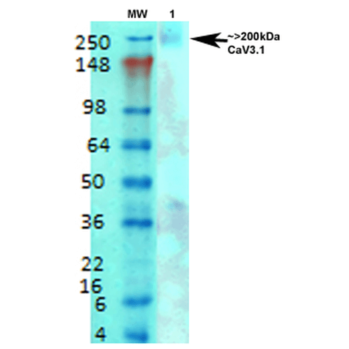 Western Blot analysis of Rat brain membrane lysate showing detection of Cav3.1 Calcium Channel protein using Mouse Anti-Cav3.1 Calcium Channel Monoclonal Antibody, Clone S178A-9 (11565). Primary Antibody: Mouse Anti-Cav3.1 Calcium Channel Monoclonal Antibody (11565) at 1:1000.
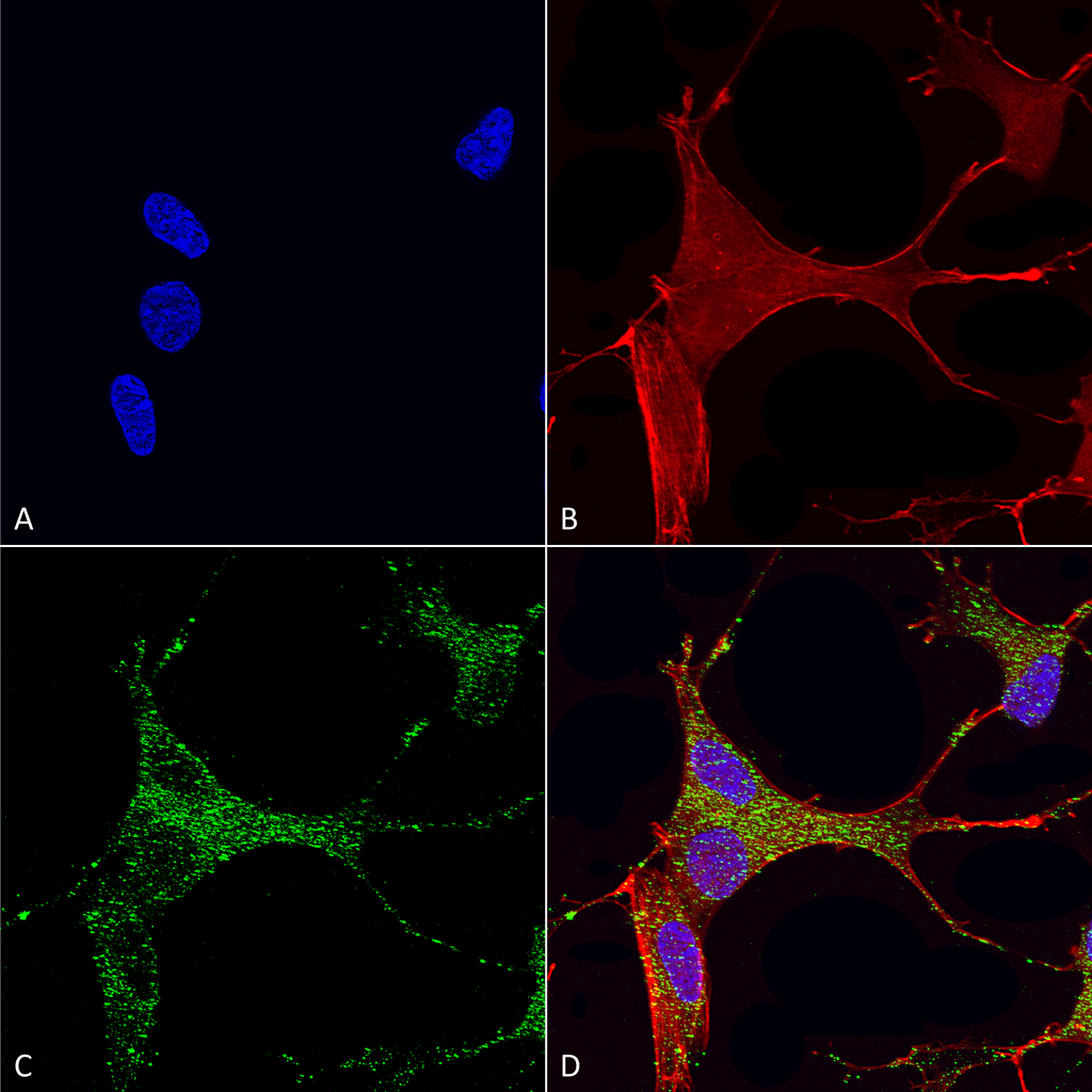 Immunocytochemistry/Immunofluorescence analysis using Mouse Anti-Cav3.1 Monoclonal Antibody, Clone S178A-9 (11565). Tissue: Neuroblastoma cells (SH-SY5Y). Species: Human. Fixation: 4% PFA for 15 min. Primary Antibody: Mouse Anti-Cav3.1 Monoclonal Antibody (11565) at 1:50 for overnight at 4°C with slow rocking. Secondary Antibody: AlexaFluor 488 at 1:1000 for 1 hour at RT. Counterstain: Phalloidin-iFluor 647 (red) F-Actin stain; Hoechst (blue) nuclear stain at 1:800, 1.6mM for 20 min at RT. (A) Hoechst (blue) nuclear stain. (B) Phalloidin-iFluor 647 (red) F-Actin stain. (C) Cav3.1 Antibody (D) Composite.
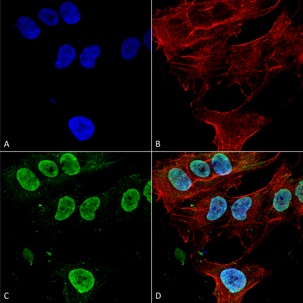 Immunocytochemistry/Immunofluorescence analysis using Mouse Anti-Cav3.1 Monoclonal Antibody, Clone S178A-9 (11565). Tissue: Neuroblastoma cell line (SK-N-BE). Species: Human. Fixation: 4% Formaldehyde for 15 min at RT. Primary Antibody: Mouse Anti-Cav3.1 Monoclonal Antibody (11565) at 1:100 for 60 min at RT. Secondary Antibody: Goat Anti-Mouse ATTO 488 at 1:200 for 60 min at RT. Counterstain: Phalloidin Texas Red F-Actin stain; DAPI (blue) nuclear stain at 1:1000, 1:5000 for 60 min at RT, 5 min at RT. Localization: Cell Membrane, Membrane, Cytoplasm, Nucleoplasm. Magnification: 60X. (A) DAPI (blue) nuclear stain. (B) Phalloidin Texas Red F-Actin stain. (C) Cav3.1 Antibody. (D) Composite.
 - -
- -
Antibody DetailsProduct DetailsReactivity Species Human ⋅ Mouse ⋅ Rat Host Species Mouse Immunogen Fusion protein corresponding to aa 2052-2172 (cytoplasmic C-terminus) of mouse Cav3.1 (accession no. NP_001106284.1). Product Concentration Lot Specific Formulation PBS, pH 7.4; 50% glycerol, 0.09% sodium azide. State of Matter Liquid Product Preparation Purified by Protein G affinity chromatography Storage and Handling This antibody is stable for at least one (1) year at -20°C. Avoid repeated freeze-thaw cycles. Regulatory Status For in vitro investigational use only. Not for use in therapeutic or diagnostic procedures. Country of Origin USA Shipping Next Day 2-8°C Applications and Recommended Usage? Quality Tested by Leinco Immunoblotting: use at 1-2ug/mL. A band of >200kDa is detected.
Immunohistochemistry: use at 1-10ug/mL. These are recommended concentrations. User should determine optimal concentrations for their application. Positive control: Rat brain membranes. Each investigator should determine their own optimal working dilution for specific applications. See directions on lot specific datasheets, as information may periodically change. DescriptionSpecificity This antibody recognizes human, mouse, and rat Cav3.1. It does not cross-react with Cav3.2. Background Ion channels are integral membrane proteins that help establish and control the small voltage gradient across the plasma membrane of living cells by allowing the flow of ions down their electrochemical gradient. Cav3.1 is a low-voltage- activated T-type calcium channel expressed throughout the body. Drugs that block T-type calcium channels are used as antihypertensives and antiepileptics and possibly in some anesthetics and antipsychotics. Antigen DetailsNCBI Gene Bank ID UniProt.org Research Area Ion Channels References & CitationsTechnical ProtocolsICC IF   |
Formats Available
 Products are for research use only. Not for use in diagnostic or therapeutic procedures.