

Anti-Fibroblast Growth Factor 13 (FGF13) [Clone S235-22]


Anti-Fibroblast Growth Factor 13 (FGF13) [Clone S235-22]
Product No.: 30109
- -
- -
Clone S235-22 Target Fibroblast Growth Factor 13 (FGF13) Formats AvailableView All Product Type Monoclonal Alternate Names FGF-13, Fibroblast growth factor homologous factor 2, FHF-2 Isotype Mouse IgG2b Applications ICC , IF , IHC , WB |
Data
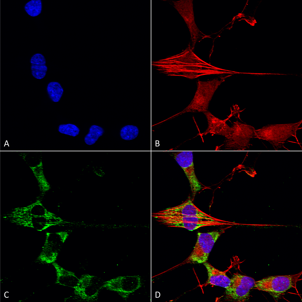 Immunocytochemistry/Immunofluorescence analysis using Mouse Anti-FGFA/FHFA (pan) Monoclonal Antibody, Clone S235-22 (30109). Tissue: Neuroblastoma cells (SH-SY5Y). Species: Human. Fixation: 4% PFA for 15 min. Primary Antibody: Mouse Anti-FGFA/FHFA (pan) Monoclonal Antibody (30109) at 1:50 for overnight at 4°C with slow rocking. Secondary Antibody: AlexaFluor 488 at 1:1000 for 1 hour at RT. Counterstain: Phalloidin-iFluor 647 (red) F-Actin stain; Hoechst (blue) nuclear stain at 1:800, 1.6mM for 20 min at RT. (A) Hoechst (blue) nuclear stain. (B) Phalloidin-iFluor 647 (red) F-Actin stain. (C) FGFA/FHFA (pan) Antibody (D) Composite.
 Immunocytochemistry/Immunofluorescence analysis using Mouse Anti-FGFA/FHFA (pan) Monoclonal Antibody, Clone S235-22 (30109). Tissue: Neuroblastoma cell line (SK-N-BE). Species: Human. Fixation: 4% Formaldehyde for 15 min at RT. Primary Antibody: Mouse Anti-FGFA/FHFA (pan) Monoclonal Antibody (30109) at 1:100 for 60 min at RT. Secondary Antibody: Goat Anti-Mouse ATTO 488 at 1:100 for 60 min at RT. Counterstain: Phalloidin Texas Red F-Actin stain; DAPI (blue) nuclear stain at 1:1000; 1:5000 for 60 min RT, 5 min RT. Localization: Cell Projection, Nucleus, Cytoplasm. Magnification: 60X. (A) DAPI (blue) nuclear stain. (B) Phalloidin Texas Red F-Actin stain. (C) FGFA/FHFA (pan) Antibody. (D) Composite.
 Western Blot analysis of Rat Brain Membrane showing detection of ~30 kDa FGFA/FHFA (pan) protein using Mouse Anti-FGFA/FHFA (pan) Monoclonal Antibody, Clone S235-22 (30109). Lane 1: Molecular Weight Ladder. Lane 2: Rat Brain Membrane. Load: 15 µg. Block: 2% BSA and 2% Skim Milk in 1X TBST. Primary Antibody: Mouse Anti-FGFA/FHFA (pan) Monoclonal Antibody (30109) at 1:200 for 16 hours at 4°C. Secondary Antibody: Goat Anti-Mouse IgG: HRP at 1:1000 for 1 hour RT. Color Development: ECL solution for 6 min in RT. Predicted/Observed Size: ~30 kDa.
 - -
- -
Antibody DetailsProduct DetailsReactive Species Human ⋅ Mouse ⋅ Rat Host Species Mouse Immunogen Fusion protein corresponding to aa 2-18 (AAAIASSLIRQKRQARE) of human FGF13. This sequence is 100 Product Concentration 1.0 mg/ml Formulation PBS, pH 7.4, 0.1% sodium azide, 50% glycerol. State of Matter Liquid Product Preparation Purified by Protein G affinity chromatography Storage and Handling This product is stable for at least one (1) year at -20°C. Regulatory Status For in vitro investigational use only. Not intended for therapeutic or diagnostic procedures. Country of Origin USA Shipping Next Day 2-8°C Applications and Recommended Usage? Quality Tested by Leinco Immunoblotting: use at 1-5ug/mL. A band of ~30kDa is detected.
Immunofluorescence: use at 10ug/mL. These are recommended concentrations. Endusers should determine optimal concentrations for their application. Each investigator should determine their own optimal working dilution for specific applications. See directions on lot specific datasheets, as information may periodically change. DescriptionDescriptionSpecificity This antibody recognizes human, mouse, and rat FGF13. Background FGF13 (also known as FGF2 and FHF2) is a member of the fibroblast growth factor family. FGF family members possess broad mitogenic and cell survival activities, and are involved in a variety of biological processes including embryonic development, cell growth, morphogenesis, tissue repair, tumor growth, and invasion. The FGF13 gene is located in a region on chromosome X that is associated with Borjeson-Forssman-Lehmann syndrome (BFLS), making it a candidate gene for familial cases of BFLS and other syndromal and nonspecific forms of X-linked mental retardation mapping to this region. Alternative splicing of this gene at the 5′ end results in several transcript variants encoding different isoforms with different N-termini. Function Microtubule-binding protein which directly binds tubulin and is involved in both polymerization and stabilization of microtubules (By similarity). Through its action on microtubules, may participate in the refinement of axons by negatively regulating axonal and leading processes branching (By similarity). Plays a crucial role in neuron polarization and migration in the cerebral cortex and the hippocampus (By similarity). Regulates voltage-gated sodium channels transport and function (PubMed:15282281, PubMed:33245860). May also play a role in MAPK signaling (By similarity). Required for the development of axonal initial segment-targeting inhibitory GABAergic synapses made by chandelier neurons (By similarity). {UniProtKB:P70377, PubMed:15282281, PubMed:33245860}. NCBI Gene Bank ID UniProt.org Research Area Growth Factors, Cytokines, Receptors References & CitationsRiesselman M et al. 2007 J Immunol 179: 2520-2531.Maaty WS et al. 2013 J Biol Chem 288: 27042-27058. Technical ProtocolsICC IF   Certificate of Analysis |
Formats Available
 Products are for research use only. Not for use in diagnostic or therapeutic procedures.