

Anti-HSP70 (HSC70) [Clone BB70]


Anti-HSP70 (HSC70) [Clone BB70]
Product No.: 11099
- -
- -
Clone BB70 Target Hsp70/Hsc70 Formats AvailableView All Product Type Monoclonal Alternate Names HSP70 Isotype Mouse IgG2a Applications ICC , IF , IHC , IP , WB |
Data
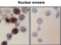 Immunocytochemistry/Immunofluorescence analysis using Mouse Anti-Hsp70 Monoclonal Antibody, Clone BB70 (11099). Tissue: hepatocyte nuclei. Species: Rat. Primary Antibody: Mouse Anti-Hsp70 Monoclonal Antibody (11099) at 1:200. Liver sections were paraffin embedded. First pictures in series show two hours after exposure to stress, the second shows the control. Courtesy of: G. Matic, University of Belgrade, Serbia.
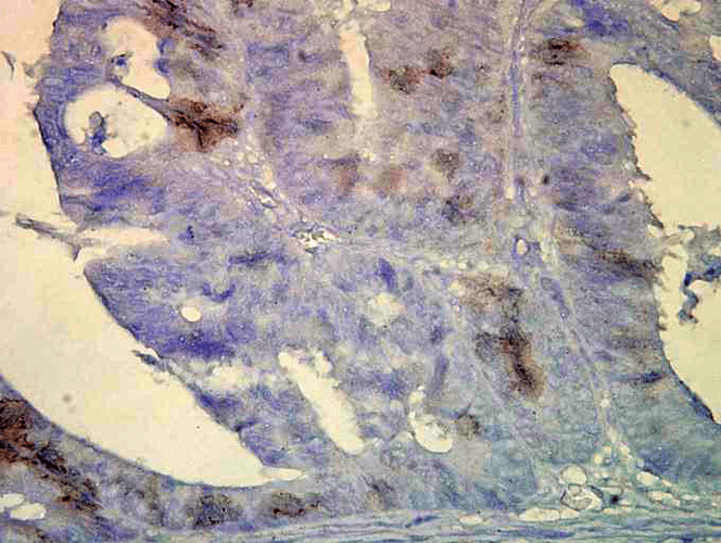 Immunohistochemistry analysis using Mouse Anti-Hsp70 Monoclonal Antibody, Clone BB70 (11099). Tissue: colon carcinoma. Species: Human. Fixation: Formalin. Primary Antibody: Mouse Anti-Hsp70 Monoclonal Antibody (11099) at 1:10000 for 12 hours at 4°C. Secondary Antibody: Biotin Goat Anti-Mouse at 1:2000 for 1 hour at RT. Counterstain: Mayer Hematoxylin (purple/blue) nuclear stain at 200 µl for 2 minutes at RT. Localization: Inflammatory cells. Magnification: 40x. HSP70/HSC70 cells stained brown. This image was produced using an amplifying IHC wash buffer. The antibody has therefore been diluted more than is recommended for other applications.
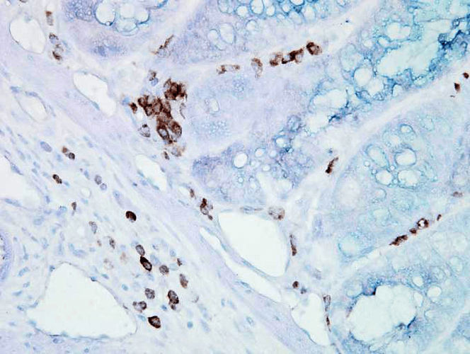 Immunohistochemistry analysis using Mouse Anti-Hsp70 Monoclonal Antibody, Clone BB70 (11099). Tissue: inflamed colon. Species: Mouse. Fixation: Formalin. Primary Antibody: Mouse Anti-Hsp70 Monoclonal Antibody (11099) at 1:10000 for 12 hours at 4°C. Secondary Antibody: Biotin Goat Anti-Mouse at 1:2000 for 1 hour at RT. Counterstain: Mayer Hematoxylin (purple/blue) nuclear stain at 200 µl for 2 minutes at RT. Localization: Inflammatory cells. Magnification: 40x. Inflammatory cells. HSP70/HSC70 stained brown. This image was produced using an amplifying IHC wash buffer. The antibody has therefore been diluted more than is recommended for other applications.
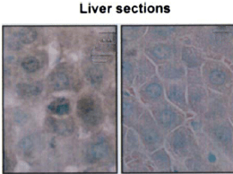 Immunohistochemistry analysis using Mouse Anti-Hsp70 Monoclonal Antibody, Clone BB70 (11099). Tissue: hepatocytes. Species: Rat. Fixation: Paraffin Embedded. Primary Antibody: Mouse Anti-Hsp70 Monoclonal Antibody (11099) at 1:200. Liver sections were paraffin embedded. First pictures in series show two hours after exposure to stress, the second shows the control. Courtesy of: G. Matic, University of Belgrade, Serbia.
 - -
- -
Antibody DetailsProduct DetailsReactivity Species Bovine ⋅ Canine ⋅ Chicken ⋅ Fish ⋅ Guinea Pig ⋅ Hamster ⋅ Human ⋅ Monkey ⋅ Mouse ⋅ Pig ⋅ Rabbit ⋅ Rat ⋅ Sheep ⋅ Xenopus ⋅ Yeast Host Species Mouse Immunogen Chicken Hsp70/Hsp90 complex. Product Concentration Lot Specific Formulation PBS, pH 7.4, 0.1% sodium azide. State of Matter Liquid Product Preparation Purified by Protein G affinity chromatography Storage and Handling This antibody is stable for at least one (1) year at -20°C. Avoid repeated freezing and thawing. Regulatory Status For in vitro investigational use only. Not for use in therapeutic or diagnostic procedures. Country of Origin USA Shipping Next Day 2-8°C Applications and Recommended Usage? Quality Tested by Leinco Immunoblotting: use at 1ug/mL.
Immunohistochemistry: use at 1-5ug/mL. These are recommended concentrations. Endusers should determine optimal concentrations for their applications. Each investigator should determine their own optimal working dilution for specific applications. See directions on lot specific datasheets, as information may periodically change. DescriptionSpecificity This antibody detects 72 and 73kDa proteins corresponding to the predicted
molecular masses of Hsp70 and Hsc70, respectively, on immunoblots of human, mouse, rat,
rabbit, monkey, hamster, guinea pig, dog, bovine, sheep, pig, chicken yeast, fish, and Xenopus
samples. This antibody recognizes the inducible and constitutive forms of Hsp70 and does not
cross-react with Hsp90. Background Members of the Hsp70 family are molecular chaperones that function in protein folding, transport, maturation and degradation by binding to nascent polypeptide chains as well as partially folded protein intermediates and preventing their aggregation and misfolding. The 70kDa heat shock cognate protein, Hsc70, is closely related biochemically and biologically to Hsp70. Antigen DetailsFunction Molecular chaperone implicated in a wide variety of cellular processes, including protection of the proteome from stress, folding and transport of newly synthesized polypeptides, activation of proteolysis of misfolded proteins and the formation and dissociation of protein complexes. Plays a pivotal role in the protein quality control system, ensuring the correct folding of proteins, the re-folding of misfolded proteins and controlling the targeting of proteins for subsequent degradation. This is achieved through cycles of ATP binding, ATP hydrolysis and ADP release, mediated by co-chaperones. The affinity for polypeptides is regulated by its nucleotide bound state. In the ATP-bound form, it has a low affinity for substrate proteins. However, upon hydrolysis of the ATP to ADP, it undergoes a conformational change that increases its affinity for substrate proteins. It goes through repeated cycles of ATP hydrolysis and nucleotide exchange, which permits cycles of substrate binding and release. {UniProtKB:P0DMV8}. NCBI Gene Bank ID UniProt.org Research Area Heat Shock & Stress Proteins References & CitationsBarent RL et al. 1998 Mol Endocrinol 12: 342. Felts SJ et al. 2000 J Biol Chem 275: 3305. Arlander SJ et al. 2003 J Biol Chem 278: 52572. Technical ProtocolsICC IF    |
Formats Available
 Products are for research use only. Not for use in diagnostic or therapeutic procedures.