Anti-Hsp90 [Clone AC-16]
Anti-Hsp90 [Clone AC-16]
Product No.: 11104
- -
- -
Clone AC-16 Target Hsp90 Formats AvailableView All Product Type Monoclonal Isotype Mouse IgG2b Applications IHC , WB , AM |
Data
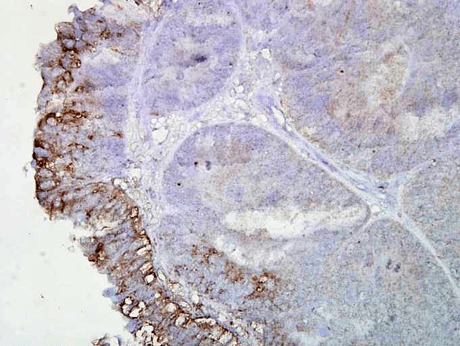
 Immunohistochemistry analysis using Mouse Anti-Hsp90 Monoclonal Antibody, Clone AC-16 (11104). Tissue: colon carcinoma. Species: Human. Fixation: Formalin. Primary Antibody: Mouse Anti-Hsp90 Monoclonal Antibody (11104) at 1:2000 for 12 hours at 4°C. Secondary Antibody: Biotin Goat Anti-Mouse at 1:2000 for 1 hour at RT. Counterstain: Mayer Hematoxylin (purple/blue) nuclear stain at 200 µl for 2 minutes at RT. Localization: Inflammatory cells. Magnification: 40x. Inflammatory cells. This image was produced using an amplifying IHC wash buffer. The antibody has therefore been diluted more than is recommended for other applications.
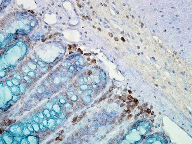
 Immunohistochemistry analysis using Mouse Anti-Hsp90 Monoclonal Antibody, Clone AC-16 (11104). Tissue: inflamed colon. Species: Mouse. Fixation: Formalin. Primary Antibody: Mouse Anti-Hsp90 Monoclonal Antibody (11104) at 1:2000 for 12 hours at 4°C. Secondary Antibody: Biotin Goat Anti-Mouse at 1:2000 for 1 hour at RT. Counterstain: Mayer Hematoxylin (purple/blue) nuclear stain at 200 µl for 2 minutes at RT. Localization: Inflammatory cells. Magnification: 40x. Mostly inflammatory cells, some mucosa. This image was produced using an amplifying IHC wash buffer. The antibody has therefore been diluted more than is recommended for other applications.
 - -
- -
Antibody DetailsProduct DetailsReactivity Species Achlya ⋅ Chicken ⋅ Human ⋅ Mouse ⋅ Rabbit ⋅ Rat ⋅ Sf9 Cells ⋅ Wheat Germ Host Species Mouse Immunogen Hsp90 purified from the water mold Achlya ambisexualis. Product Concentration Lot Specific Formulation PBS, pH 7.4. State of Matter Liquid Product Preparation Purified by Protein G affinity chromatography Storage and Handling This antibody is stable for at least one (1) year at -20°C. Avoid repeated freezing and
thawing. Regulatory Status For in vitro investigational use only. Not for
use in therapeutic or diagnostic procedures.m Country of Origin USA Shipping Next Day 2-8°C Applications and Recommended Usage? Quality Tested by Leinco Immunoblotting: use at 1ug/mL. A band of ~88 kDa is detected.
These are recommended concentrations. User should determine optimal concentrations for their application. Positive control: Heat shocked HeLa cell lysate. Each investigator should determine their own optimal working dilution for specific applications. See directions on lot specific datasheets, as information may periodically change. DescriptionSpecificity This antibody is reactive with both the
constitutive and the inducible forms of
human, mouse, rat, rabbit, chicken, Sf9 cell,
Achlya, and wheat germ Hsp90. However, it
does not bind to the native form of Hsp90
and does not recognize E. coli and yeast
Hsp90. Background Hsp90 is a highly conserved and essential stress protein that is expressed in all eukaryotic cells. It participates in the folding, assembly, maturation, and stabilization of specific proteins as an integral component of chaperone complexes. Hsp90 is relatively abundant in unstressed cells of most prokaryotic and eukaryotic systems and can be induced by heat shock in some systems. It exists in a dimeric form and has been observed to bind to several other cellular proteins such as retrovirus kinases, steroid receptors, hemeregulated protein kinase, actin, and tubulin. When bound to ATP, Hsp 90 interacts with co-chaperones cdc37, p23, and various immunophilin-like proteins, forming complexes that stabilize and protect target proteins from proteasomal degradation. Antigen DetailsNCBI Gene Bank ID UniProt.org Research Area Heat Shock & Stress Proteins References & CitationsTechnical Protocols |


