

Anti-Leucine-Rich Glioma Inactivated Gene 1 (LGI1) Antibody (56577)


Anti-Leucine-Rich Glioma Inactivated Gene 1 (LGI1) Antibody (56577)
Product No.: 56577
- -
- -
Clone S283-7 Target Leucine-Rich Glioma Inactivated Gene 1 (LGI1) Formats AvailableView All Product Type Monoclonal Isotype Mouse IgG2a Applications ICC , IF , IHC , WB |
Data
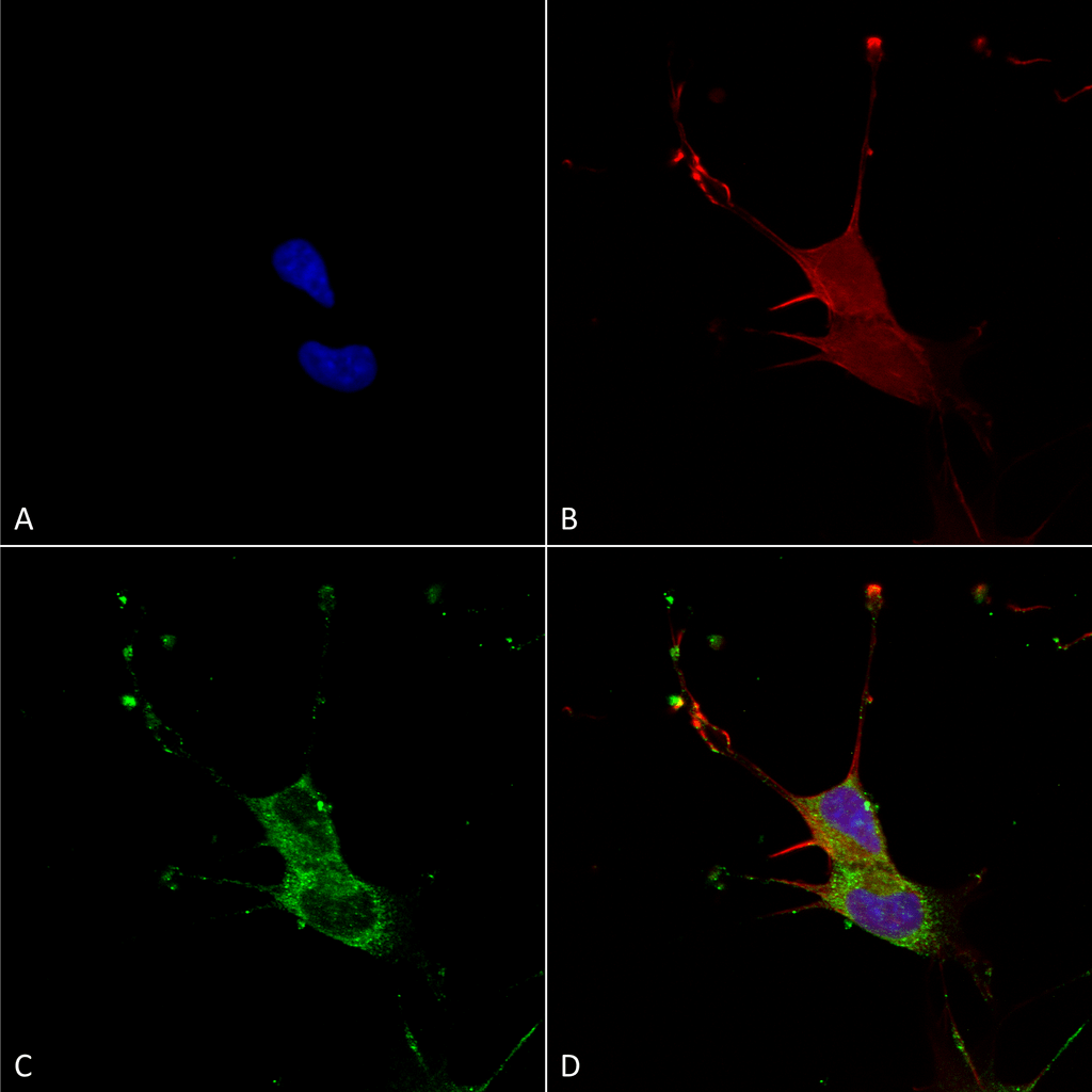 Immunocytochemistry/Immunofluorescence analysis using Mouse Anti-LGI1 Monoclonal Antibody, Clone S283-7 (56577). Tissue: Neuroblastoma cells (SH-SY5Y). Species: Human. Fixation: 4% PFA for 15 min. Primary Antibody: Mouse Anti-LGI1 Monoclonal Antibody (56577) at 1:100 for overnight at 4°C with slow rocking. Secondary Antibody: AlexaFluor 488 at 1:1000 for 1 hour at RT. Counterstain: Phalloidin-iFluor 647 (red) F-Actin stain; Hoechst (blue) nuclear stain at 1:800, 1.6mM for 20 min at RT. (A) Hoechst (blue) nuclear stain. (B) Phalloidin-iFluor 647 (red) F-Actin stain. (C) LGI1 Antibody (D) Composite.
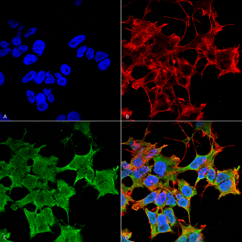 Immunocytochemistry/Immunofluorescence analysis using Mouse Anti-LGI1 Monoclonal Antibody, Clone S283-7 (56577). Tissue: Neuroblastoma cell line (SK-N-BE). Species: Human. Fixation: 4% Formaldehyde for 15 min at RT. Primary Antibody: Mouse Anti-LGI1 Monoclonal Antibody (56577) at 1:100 for 60 min at RT. Secondary Antibody: Goat Anti-Mouse ATTO 488 at 1:100 for 60 min at RT. Counterstain: Phalloidin Texas Red F-Actin stain; DAPI (blue) nuclear stain at 1:1000, 1:5000 for 60min RT, 5min RT. Localization: Cell Junction, Golgi Apparatus, Endoplasmic Reticulum. Magnification: 60X. (A) DAPI (blue) nuclear stain. (B) Phalloidin Texas Red F-Actin stain. (C) LGI1 Antibody. (D) Composite.
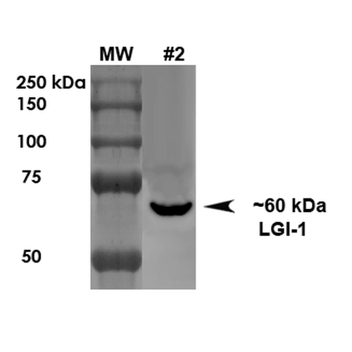 Western Blot analysis of Rat Brain Membrane showing detection of ~60 kDa LGI1 protein using Mouse Anti-LGI1 Monoclonal Antibody, Clone S283-7 (56577). Load: 10 µg. Primary Antibody: Mouse Anti-LGI1 Monoclonal Antibody (56577) at 1:1000 for 1 hour at RT. Secondary Antibody: Goat Anti-Mouse HRP at 1:200 for 1 hour at RT. Predicted/Observed Size: ~60 kDa.
 - -
- -
Antibody DetailsProduct DetailsReactive Species Human ⋅ Mouse ⋅ Rat Host Species Mouse Immunogen Fusion protein corresponding to aa 37-113 of mouse LGI1. This sequence is 100 Product Concentration 1.0 mg/ml Formulation PBS, pH 7.4, 0.1% sodium azide, 50% glycerol. State of Matter Liquid Product Preparation Purified by Protein G affinity chromatography Storage and Handling This product is stable for at least one (1) year at -20°C. Regulatory Status For in vitro investigational use only. Not intended for therapeutic or diagnostic procedures. Country of Origin USA Shipping Next Day 2-8°C Applications and Recommended Usage? Quality Tested by Leinco Immunoblotting: use at 1-5ug/mL. A band of ~60kDa is detected.
Immunofluorescence: use at 10ug/mL. These are recommended concentrations. Endusers should determine optimal concentrations for their application. Each investigator should determine their own optimal working dilution for specific applications. See directions on lot specific datasheets, as information may periodically change. DescriptionDescriptionSpecificity This antibody recognizes human, mouse, and rat LGI1. Background The leucine-rich glioma inactivated -1 (LGI1) gene is rearranged as a result of translocations in glioblastoma cell lines. This protein contains a hydrophobic segment representing a putative transmembrane domain with the amino terminus located outside the cell. It also contains leucine- rich repeats with conserved cysteine-rich flanking sequences. This gene is predominantly expressed in neural tissues and its expression is reduced in low grade brain tumors and significantly reduced or absent in malignant gliomas. NCBI Gene Bank ID UniProt.org Research Area Neuroscience References & CitationsTechnical ProtocolsICC IF   Certificate of Analysis |
Formats Available
- -
- -
Prod No. | Description |
|---|---|
56577 |
 Products are for research use only. Not for use in diagnostic or therapeutic procedures.