

Anti-Mitofusin-2 [Clone S153-5]


Anti-Mitofusin-2 [Clone S153-5]
Product No.: 56557
- -
- -
Clone S153-5 Target Mitofusin-2 Formats AvailableView All Product Type Monoclonal Alternate Names EC 3.6.5.-, Hypertension-related protein 1, Mitochondrial assembly regulatory factor, HSG protein, Transmembrane GTPase MFN2 Isotype Mouse IgG2a Applications ICC , IF , IHC , WB |
Data
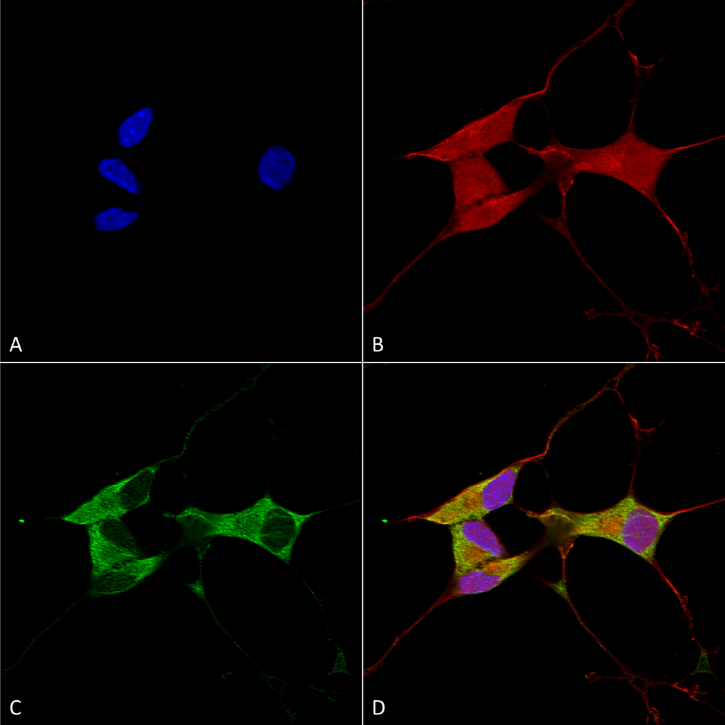 Immunocytochemistry/Immunofluorescence analysis using Mouse Anti-Mitofusin 2 Monoclonal Antibody, Clone S153-5 (56557). Tissue: Neuroblastoma cells (SH-SY5Y). Species: Human. Fixation: 4% PFA for 15 min. Primary Antibody: Mouse Anti-Mitofusin 2 Monoclonal Antibody (56557) at 1:100 for overnight at 4°C with slow rocking. Secondary Antibody: AlexaFluor 488 at 1:1000 for 1 hour at RT. Counterstain: Phalloidin-iFluor 647 (red) F-Actin stain; Hoechst (blue) nuclear stain at 1:800, 1.6mM for 20 min at RT. (A) Hoechst (blue) nuclear stain. (B) Phalloidin-iFluor 647 (red) F-Actin stain. (C) Mitofusin 2 Antibody (D) Composite.
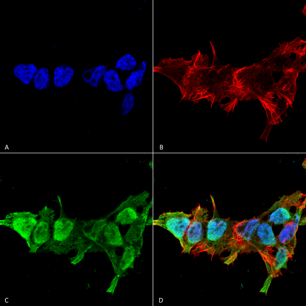 Immunocytochemistry/Immunofluorescence analysis using Mouse Anti-Mitofusin 2 Monoclonal Antibody, Clone S153-5 (56557). Tissue: Neuroblastoma cell line (SK-N-BE). Species: Human. Fixation: 4% Formaldehyde for 15 min at RT. Primary Antibody: Mouse Anti-Mitofusin 2 Monoclonal Antibody (56557) at 1:100 for 60 min at RT. Secondary Antibody: Goat Anti-Mouse ATTO 488 at 1:100 for 60 min at RT. Counterstain: Phalloidin Texas Red F-Actin stain; DAPI (blue) nuclear stain at 1:1000; 1:5000 for 60 min RT, 5 min RT. Localization: Cytoplasm, Nucleus. Magnification: 60X. (A) DAPI (blue) nuclear stain. (B) Phalloidin Texas Red F-Actin stain. (C) Mitofusin 2 Antibody. (D) Composite.
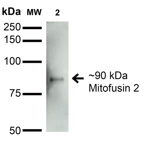 Western Blot analysis of Rat Brain Membrane showing detection of ~90 kDa Mitofusin 2 protein using Mouse Anti-Mitofusin 2 Monoclonal Antibody, Clone S153-5 (56557). Lane 1: Molecular Weight Ladder. Lane 2: Rat Brain Membrane. Load: 15 µg. Block: 2% BSA and 2% Skim Milk in 1X TBST. Primary Antibody: Mouse Anti-Mitofusin 2 Monoclonal Antibody (56557) at 1:200 for 16 hours at 4°C. Secondary Antibody: Goat Anti-Mouse IgG: HRP at 1:1000 for 1 hour RT. Color Development: ECL solution for 6 min in RT. Predicted/Observed Size: ~90 kDa.
 - -
- -
Antibody DetailsProduct DetailsReactive Species Human ⋅ Mouse ⋅ Rat Host Species Mouse Immunogen Fusion protein corresponding to aa 370-600 (cytoplasmic N-terminus) of mouse Mitofusin-2. This sequence is 97 Product Concentration 1.0 mg/ml Formulation PBS, pH 7.4, 0.1% sodium azide, 50% glycerol. State of Matter Liquid Product Preparation Purified by Protein G affinity chromatography Storage and Handling This antibody is stable for at least one (1) year at -20°C. Avoid multiple freeze-thaw cycles. Regulatory Status For in vitro investigational use only. Not intended for therapeutic or diagnostic procedures. Country of Origin USA Shipping Next Day 2-8°C Applications and Recommended Usage? Quality Tested by Leinco Immunoblotting: use at 1-5ug/mL. A band of ~90kDa is detected.
Immunofluorescence: use at 10µg/mL. These are recommended concentrations. Enduser should determine optimal concentrations for their application. Each investigator should determine their own optimal working dilution for specific applications. See directions on lot specific datasheets, as information may periodically change. DescriptionDescriptionSpecificity This antibody recognizes human, mouse, and rat Mitofusin 2. It does not cross-react with Mitofusin 1. Background Mitofusin-2 (Mfn2) is a mitochondrial membrane protein that participates in mitochondrial fusion and contributes to the maintenance and operation of the mitochondrial network. This protein is involved in the regulation of vascular smooth muscle cell proliferation, and it may play a role in the pathophysiology of obesity. Mutations in this gene cause Charcot-Marie-Tooth disease type 2A2 and hereditary motor and sensory neuropathy VI, both of which are disorders of the peripheral nervous system. Defects in this gene have also been associated with early-onset stroke. Two transcript variants encoding the same protein have been identified. Function Mitochondrial outer membrane GTPase that mediates mitochondrial clustering and fusion (PubMed:12527753, PubMed:23921378, PubMed:23620051). Mitochondria are highly dynamic organelles, and their morphology is determined by the equilibrium between mitochondrial fusion and fission events. Overexpression induces the formation of mitochondrial networks. Membrane clustering requires GTPase activity and may involve a major rearrangement of the coiled coil domains (By similarity). Plays a central role in mitochondrial metabolism and may be associated with obesity and/or apoptosis processes. Plays an important role in the regulation of vascular smooth muscle cell proliferation (By similarity). Involved in the clearance of damaged mitochondria via selective autophagy (mitophagy). Is required for PRKN recruitment to dysfunctional mitochondria (PubMed:23620051). Involved in the control of unfolded protein response (UPR) upon ER stress including activation of apoptosis and autophagy during ER stress (PubMed:23921556). Acts as an upstream regulator of EIF2AK3 and suppresses EIF2AK3 activation under basal conditions (PubMed:23921556). {UniProtKB:O95140, UniProtKB:Q8R500, PubMed:12527753, PubMed:23620051, PubMed:23921378, PubMed:23921556}. NCBI Gene Bank ID UniProt.org Research Area Neuroscience References & CitationsTechnical ProtocolsICC IF   Certificate of Analysis |
Formats Available
 Products are for research use only. Not for use in diagnostic or therapeutic procedures.