

Anti-Mitosin Antibody (3320)


Anti-Mitosin Antibody (3320)
Product No.: 3320
- -
- -
Clone 14C10 Target Mitosin Formats AvailableView All Product Type Monoclonal Alternate Names CENP-F, AH antigen, Kinetochore protein CENPF, Mitosin Isotype Mouse IgG2a Applications FACS , ICC , IF , IHC FF , IHC FFPE , IP , WB |
Data
![CENPF antibody [14C10 1D8] detects CENPF protein at nucleus on BT483 xenograft by immunohistochemical analysis. Sample: Paraffin-embedded BT483 xenograft. CENPF antibody [14C10 1D8] (3320) dilution: 1:200. Antigen Retrieval: Trilogy™ (EDTA based, pH 8.0) buffer, 15min](https://www.leinco.com/wp-content/uploads/2025/01/qed-bioscience-anti-mitosin-antibody-3320-1.jpg) CENPF antibody [14C10 1D8] detects CENPF protein at nucleus on BT483 xenograft by immunohistochemical analysis.
Sample: Paraffin-embedded BT483 xenograft.
CENPF antibody [14C10 1D8] (3320) dilution: 1:200.
Antigen Retrieval: Trilogy™ (EDTA based, pH 8.0) buffer, 15min
CENPF antibody [14C10 1D8] detects CENPF protein at nucleus on BT483 xenograft by immunohistochemical analysis.
Sample: Paraffin-embedded BT483 xenograft.
CENPF antibody [14C10 1D8] (3320) dilution: 1:200.
Antigen Retrieval: Trilogy™ (EDTA based, pH 8.0) buffer, 15min![CENPF antibody [14C10 1D8] detects CENPF protein by western blot analysis. A. 30 ug HeLa whole cell lysate/extract (untreated) B. 30 ug HeLa whole cell lysate/extract (100ng/ml Nocodazole treatment for 24hr) 5% SDS-PAGE CENPF antibody [14C10 1D8] (3320) dilution: 1:500 The HRP-conjugated anti-mouse IgG antibody was used to detect the primary antibody.](https://www.leinco.com/wp-content/uploads/2025/01/qed-bioscience-anti-mitosin-antibody-3320-2.jpg) CENPF antibody [14C10 1D8] detects CENPF protein by western blot analysis.
A. 30 ug HeLa whole cell lysate/extract (untreated)
B. 30 ug HeLa whole cell lysate/extract (100ng/ml Nocodazole treatment for 24hr)
5% SDS-PAGE
CENPF antibody [14C10 1D8] (3320) dilution: 1:500
The HRP-conjugated anti-mouse IgG antibody was used to detect the primary antibody.
CENPF antibody [14C10 1D8] detects CENPF protein by western blot analysis.
A. 30 ug HeLa whole cell lysate/extract (untreated)
B. 30 ug HeLa whole cell lysate/extract (100ng/ml Nocodazole treatment for 24hr)
5% SDS-PAGE
CENPF antibody [14C10 1D8] (3320) dilution: 1:500
The HRP-conjugated anti-mouse IgG antibody was used to detect the primary antibody.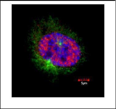 Confocal immunofluorescence staining (Olympus FV10i) of Mitosin. U2OS cells were fixed by 4% PFA and costained with Mitosin 14C10 1D8 AB (Red; cat# 3320; 1:500 Ab dilution) and alpha-tubulin rabbit polyclonal AB, a spindle marker. DAPI (blue), chromosomes. Scale bar, 5 um
 - -
- -
Antibody DetailsProduct DetailsReactive Species Human Host Species Mouse Immunogen GST fusion protein expressed in E. coli corresponding to aa 1759-2093 of human mitosin. Product Concentration Lot Specific Formulation PBS, pH 7.4 State of Matter Liquid Product Preparation Purified by Protein G affinity chromatography Storage and Handling This antibody is stable for at least one (1) year at -70°C. Avoid multiple freeze-thaw cycles. Regulatory Status Research Use Only Country of Origin USA Shipping Next Day 2-8°C Applications and Recommended Usage? Quality Tested by Leinco Immunoblotting: use at 1-10 ug/mL.
Immunoprecipitation: use at 1-10 ug/mL. Immunohistochemistry: use at 10 ug/mL with frozen or paraffin-embedded sections. Positive controls: Human tissue containing rapidly proliferating cells, such as lymphoid germinal centers. Each investigator should determine their own optimal working dilution for specific applications. See directions on lot specific datasheets, as information may periodically change. DescriptionSpecificity This antibody recognizes mitosin, a 350 kD phosphoprotein that is specifically involved in mitotic-phase progression. Mitosin is expressed throughout S, G2, and M phases of the cell cycle, but is absent in G0 and G1. It associates with the mitotic apparatus during M-phase and is redistributed from the nucleus to the centromere, spindle, and midbody during M-phase progression. Its usefulness as a proliferation marker is under active investigation. Function Required for kinetochore function and chromosome segregation in mitosis. Required for kinetochore localization of dynein, LIS1, NDE1 and NDEL1. Regulates recycling of the plasma membrane by acting as a link between recycling vesicles and the microtubule network though its association with STX4 and SNAP25. Acts as a potential inhibitor of pocket protein-mediated cellular processes during development by regulating the activity of RB proteins during cell division and proliferation. May play a regulatory or permissive role in the normal embryonic cardiomyocyte cell cycle and in promoting continued mitosis in transformed, abnormally dividing neonatal cardiomyocytes. Interaction with RB directs embryonic stem cells toward a cardiac lineage. Involved in the regulation of DNA synthesis and hence cell cycle progression, via its C-terminus. Has a potential role regulating skeletal myogenesis and in cell differentiation in embryogenesis. Involved in dendritic cell regulation of T-cell immunity against chlamydia. {PubMed:12974617, PubMed:17600710, PubMed:7542657, PubMed:7651420}. NCBI Gene Bank ID UniProt.org Research Area Cancer Research References & CitationsAllred, DC et al. (1997) Lab Invest 15A (69) Zhu, X et al. (1995), Mol Cell Biol 15: 5017-5029. Technical ProtocolsFACS ICC IF IHC FF IHC FFPE   Certificate of Analysis |
Formats Available
 Products are for research use only. Not for use in diagnostic or therapeutic procedures.