

Anti-NR2B Glutamate Receptor [Clone S59-36]


Anti-NR2B Glutamate Receptor [Clone S59-36]
Product No.: 56461
- -
- -
Clone S59-36 Target NR2B Glutamate Receptor Formats AvailableView All Product Type Monoclonal Alternate Names GluN2B, Glutamate [NMDA] receptor subunitε-2, N-methyl D-aspartate receptor subtype 2B, NMDAR2B, NR2B Isotype Mouse IgG2b Applications ICC , IF , IHC , IP , WB , AM |
Data
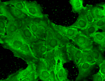 Immunocytochemistry/Immunofluorescence analysis using Mouse Anti-GluN2B/NR2B Monoclonal Antibody, Clone S59-36 (56461). Tissue: HaCaT cells. Species: Human. Fixation: Cold 100% methanol for 10 minutes at -20°C. Primary Antibody: Mouse Anti-GluN2B/NR2B Monoclonal Antibody (56461) at 1:100 for 1 hour at RT. Secondary Antibody: FITC Goat Anti-Mouse (green) at 1:50 for 1 hour at RT. Localization: Everything positive.
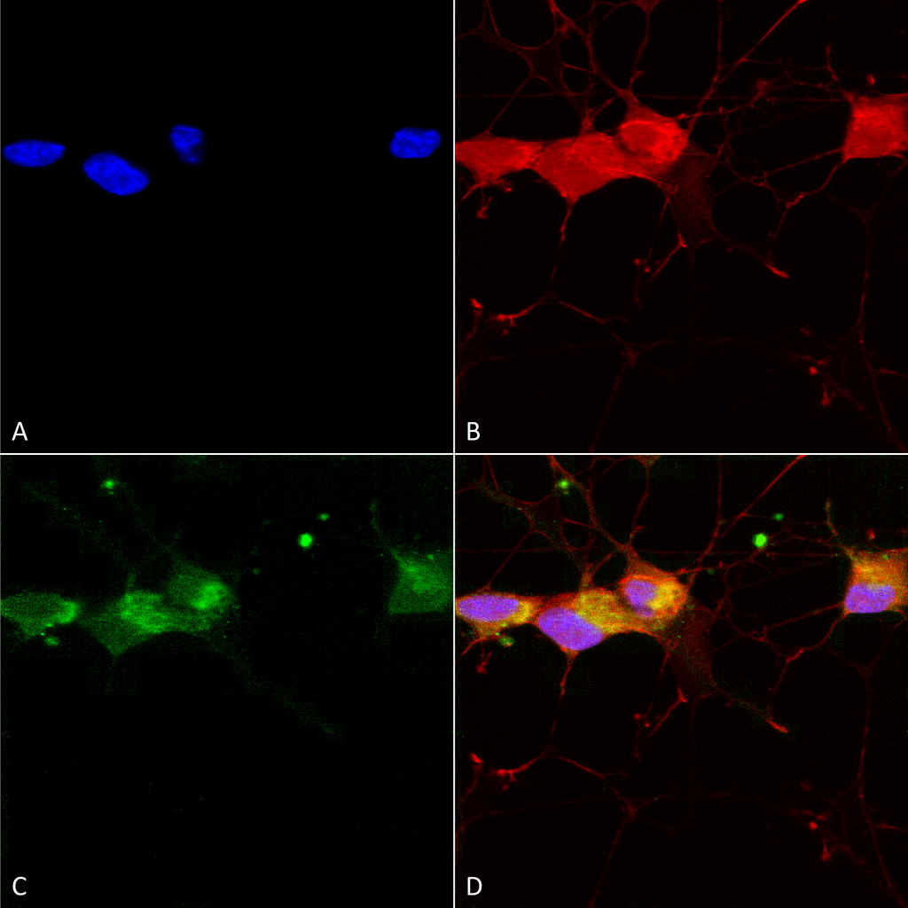 Immunocytochemistry/Immunofluorescence analysis using Mouse Anti-GluN2B/NR2B Monoclonal Antibody, Clone S59-36 (56461). Tissue: Neuroblastoma cells (SH-SY5Y). Species: Human. Fixation: 4% PFA for 15 min. Primary Antibody: Mouse Anti-GluN2B/NR2B Monoclonal Antibody (56461) at 1:50 for overnight at 4°C with slow rocking. Secondary Antibody: AlexaFluor 488 at 1:1000 for 1 hour at RT. Counterstain: Phalloidin-iFluor 647 (red) F-Actin stain; Hoechst (blue) nuclear stain at 1:800, 1.6mM for 20 min at RT. (A) Hoechst (blue) nuclear stain. (B) Phalloidin-iFluor 647 (red) F-Actin stain. (C) GluN2B/NR2B Antibody (D) Composite.
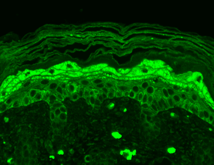 Immunohistochemistry analysis using Mouse Anti-GluN2B/NR2B Monoclonal Antibody, Clone S59-36 (56461). Tissue: backskin. Species: Mouse. Fixation: Bouin’s Fixative and paraffin-embedded. Primary Antibody: Mouse Anti-GluN2B/NR2B Monoclonal Antibody (56461) at 1:100 for 1 hour at RT. Secondary Antibody: FITC Goat Anti-Mouse (green) at 1:50 for 1 hour at RT. Localization: Filaggrin-like staining, and dermal staining.
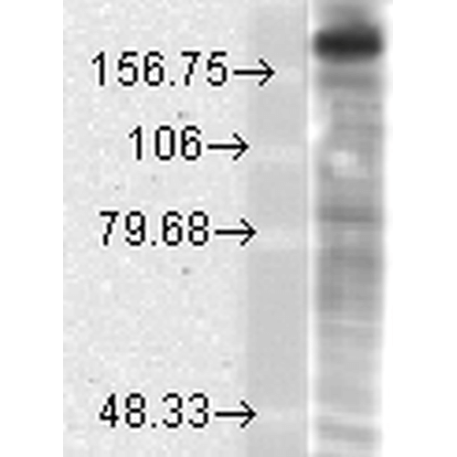 Western Blot analysis of Rat brain membrane lysate showing detection of GluN2B/NR2B protein using Mouse Anti-GluN2B/NR2B Monoclonal Antibody, Clone S59-36 (56461). Load: 15 µg. Block: 1.5% BSA for 30 minutes at RT. Primary Antibody: Mouse Anti-GluN2B/NR2B Monoclonal Antibody (56461) at 1:1000 for 2 hours at RT. Secondary Antibody: Sheep Anti-Mouse IgG: HRP for 1 hour at RT.
 - -
- -
Antibody DetailsProduct DetailsReactive Species Human ⋅ Mouse ⋅ Rat Host Species Mouse Immunogen Fusion protein aa 20-271 (extracellular N-terminus) of rat NR2B (accession no. Q00960). This sequence is 99 Product Concentration Lot Specific Formulation PBS, pH 7.4; 50% glycerol, 0.09% sodium azide. Purified by Protein G affinity chromatography. State of Matter Liquid Product Preparation Purified by Protein G affinity chromatography Storage and Handling This antibody is stable for at least one (1) year at -20°C. Avoid multiple freeze-thaw cycles. Country of Origin USA Shipping Next Day 2-8°C Applications and Recommended Usage? Quality Tested by Leinco Immunoblotting: use at 1-10ug/mL. A band of ~166kDa is detected.Immunohistochemistry and
Immunocytochemistry: use at 0.1-1ug/mL Immunofluorescence: use at 1-10ug/mL These are recommended concentrations. User should determine optimal concentrations for their application. Positive control: Rat brain lysate Each investigator should determine their own optimal working dilution for specific applications. See directions on lot specific datasheets, as information may periodically change. DescriptionDescriptionSpecificity Mouse Monoclonal Antibody specific to NR2B Glutamate Receptor Background Glutamate receptors of the NMDA subtype are involved in several physiological and pathological processes in the brain. Functional NMDA receptors are heteromultimeric complexes of the NR1 subunit and one or more of the four NR2 subunits (NR2A-D). Coexpression of both subunit types are required for the for- mation of fully functional channels that are inserted into the plasma membrane. The NR2 subunit determines characteristics of the channel such as conductance, mean open time, and sensitivity to Mg+2. The ability of NR1 and NR2 subunit combinations to flux Ca+2 is essential to the role that NMDA receptors play in synaptic plasticity and neurotoxicity. Function Component of NMDA receptor complexes that function as heterotetrameric, ligand-gated ion channels with high calcium permeability and voltage-dependent sensitivity to magnesium. Channel activation requires binding of the neurotransmitter glutamate to the epsilon subunit, glycine binding to the zeta subunit, plus membrane depolarization to eliminate channel inhibition by Mg(2+) (PubMed:1350383, PubMed:19910922, PubMed:21677647, PubMed:24876489, PubMed:27135925, PubMed:27916457). Sensitivity to glutamate and channel kinetics depend on the subunit composition (Probable). In concert with DAPK1 at extrasynaptic sites, acts as a central mediator for stroke damage. Its phosphorylation at Ser-1303 by DAPK1 enhances synaptic NMDA receptor channel activity inducing injurious Ca2+ influx through them, resulting in an irreversible neuronal death. Contributes to neural pattern formation in the developing brain. Plays a role in long-term depression (LTD) of hippocampus membrane currents and in synaptic plasticity (By similarity). {UniProtKB:Q01097, PubMed:1350383, PubMed:19910922, PubMed:21677647, PubMed:24876489, PubMed:27135925, PubMed:27916457, ECO:0000305}. NCBI Gene Bank ID UniProt.org Research Area Neuroscience References & CitationsTechnical ProtocolsICC IF    AM Certificate of Analysis |
Formats Available
 Products are for research use only. Not for use in diagnostic or therapeutic procedures.