

Anti-Protein Tyrosine Phosphatase Receptor F (PTPRF) Antibody


Anti-Protein Tyrosine Phosphatase Receptor F (PTPRF) Antibody
Product No.: 56579
- -
- -
Clone S165-38 Target Protein Tyrosine Phosphatase Receptor F (PTPRF) Formats AvailableView All Product Type Monoclonal Alternate Names EC 3.1.3.48, Leukocyte common antigen related, LAR Isotype Mouse IgG2a Applications ICC , IF , WB |
Data
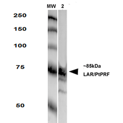 Western Blot analysis of Rat Brain Membrane showing detection of LAR protein using Mouse Anti-LAR Monoclonal Antibody, Clone S165-38 (56579). Primary Antibody: Mouse Anti-LAR Monoclonal Antibody (56579) at 1:250.
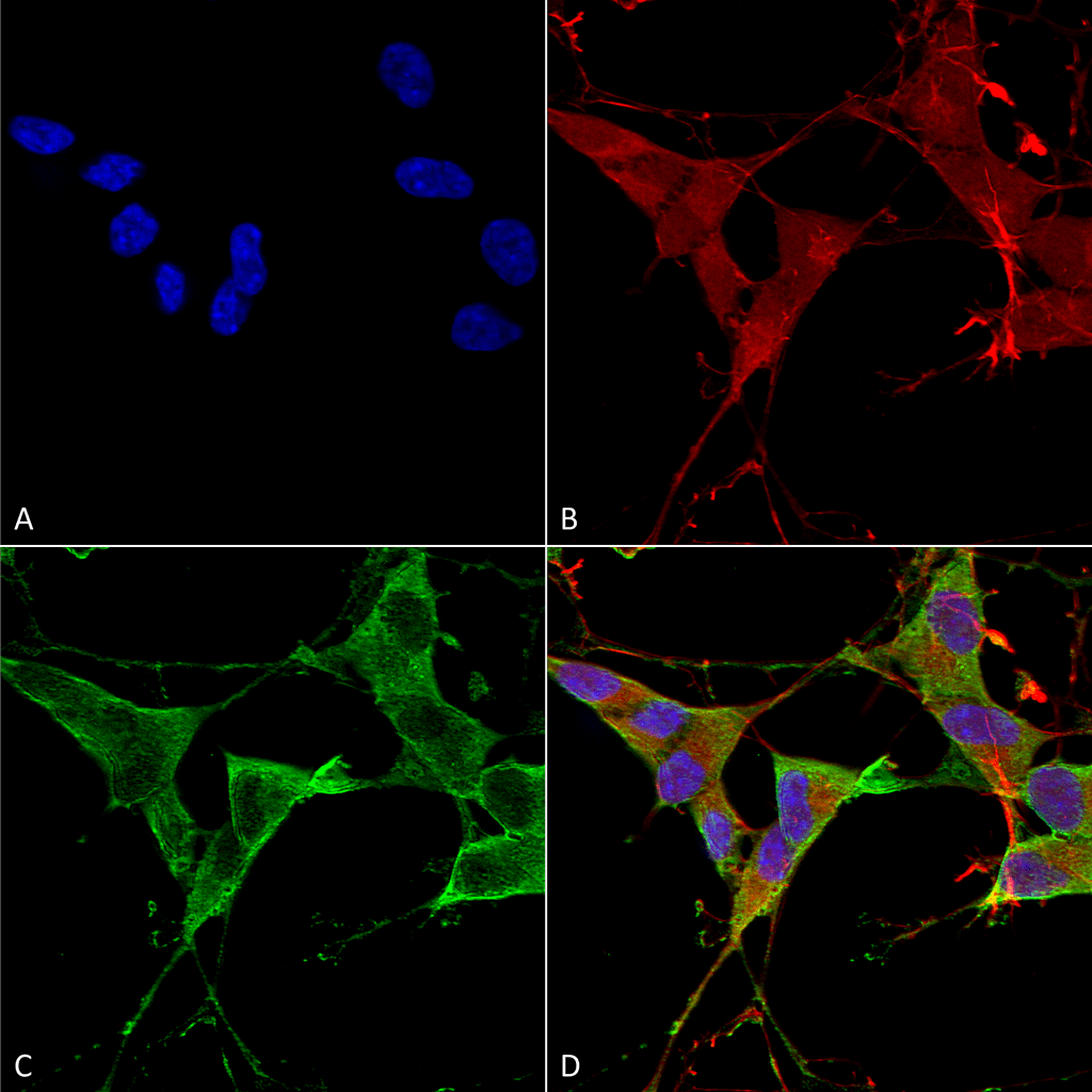 Immunocytochemistry/Immunofluorescence analysis using Mouse Anti-LAR/PTPRF Monoclonal Antibody, Clone S165-38 (56579). Tissue: Neuroblastoma cells (SH-SY5Y). Species: Human. Fixation: 4% PFA for 15 min. Primary Antibody: Mouse Anti-LAR/PTPRF Monoclonal Antibody (56579) at 1:100 for overnight at 4°C with slow rocking. Secondary Antibody: AlexaFluor 488 at 1:1000 for 1 hour at RT. Counterstain: Phalloidin-iFluor 647 (red) F-Actin stain; Hoechst (blue) nuclear stain at 1:800, 1.6mM for 20 min at RT. (A) Hoechst (blue) nuclear stain. (B) Phalloidin-iFluor 647 (red) F-Actin stain. (C) LAR/PTPRF Antibody (D) Composite.
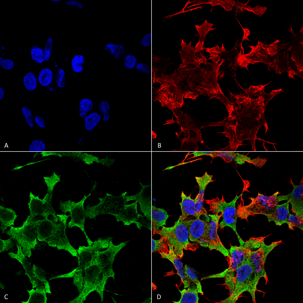 Immunocytochemistry/Immunofluorescence analysis using Mouse Anti-LAR/PTPRF Monoclonal Antibody, Clone S165-38 (56579). Tissue: Neuroblastoma cell line (SK-N-BE). Species: Human. Fixation: 4% Formaldehyde for 15 min at RT. Primary Antibody: Mouse Anti-LAR/PTPRF Monoclonal Antibody (56579) at 1:100 for 60 min at RT. Secondary Antibody: Goat Anti-Mouse ATTO 488 at 1:100 for 60 min at RT. Counterstain: Phalloidin Texas Red F-Actin stain; DAPI (blue) nuclear stain at 1:1000, 1:5000 for 60min RT, 5min RT. Localization: Membrane. Magnification: 60X. (A) DAPI (blue) nuclear stain. (B) Phalloidin Texas Red F-Actin stain. (C) LAR/PTPRF Antibody. (D) Composite.
 - -
- -
Antibody DetailsProduct DetailsReactive Species Human ⋅ Mouse ⋅ Rat Host Species Mouse Immunogen Fusion protein corresponding to aa 1315-1607 (cytoplasmic C-terminus) of human PTPRF. This sequence is 97 Product Concentration 1.0 mg/ml Formulation PBS, pH 7.4, 0.1% sodium azide, 50% glycerol. State of Matter Liquid Product Preparation Purified by Protein G affinity chromatography Storage and Handling This product is stable for at least one (1) year at -20°C. Regulatory Status For in vitro investigational use only. Not intended for therapeutic or diagnostic procedures. Country of Origin USA Shipping Next Day 2-8°C Applications and Recommended Usage? Quality Tested by Leinco Immunoblotting: use at 1-5ug/mL. A band of ~85kDa is detected which represents the subunit containing transmembrane and intracellular domains.
Immunofluorescence: use at 10ug/mL. These are recommended concentrations. Endusers should determine optimal concentrations for their application. Each investigator should determine their own optimal working dilution for specific applications. See directions on lot specific datasheets, as information may periodically change. DescriptionDescriptionSpecificity This antibody recognizes human, mouse, and rat PTPRF. Background PTPRF, like other members of the protein tyrosine phosphatase (PTP) family, is a signaling molecule that regulates a variety of cellular processes. PTPRF possesses an extracellular region, a single transmembrane region, and two tandem intracytoplasmic catalytic domains, and represents a receptor-type PTP. The extracellular region contains three Ig-like domains and nine non-Ig like domains similar to that of neural-cell adhesion molecule. PTPRF functions in the regulation of epithelial cell-cell contacts at adherents junctions as well as in the control of beta- catenin signaling. An increased expression level of PTPRF has been found in the insulin- responsive tissue of obese, insulin-resistant individuals and may contribute to the pathogenesis of insulin resistance. Two alternatively spliced transcript variants of this gene, which encode distinct proteins, have been reported. Function Possible cell adhesion receptor. It possesses an intrinsic protein tyrosine phosphatase activity (PTPase) and dephosphorylates EPHA2 regulating its activity.; The first PTPase domain has enzymatic activity, while the second one seems to affect the substrate specificity of the first one. NCBI Gene Bank ID UniProt.org Research Area Neuroscience References & CitationsTechnical ProtocolsICC IF  Certificate of Analysis |
Formats Available
 Products are for research use only. Not for use in diagnostic or therapeutic procedures.