

Anti-Rad51 Antibody (56214)


Anti-Rad51 Antibody (56214)
Product No.: 56214
- -
- -
Clone 14B4 Target Rad51 Formats AvailableView All Product Type Monoclonal Alternate Names HsRAD51, hRAD51, RAD51 homolog A Isotype Mouse IgG2b Applications ICC , IF , IHC , IHC FFPE , IP , WB |
Data
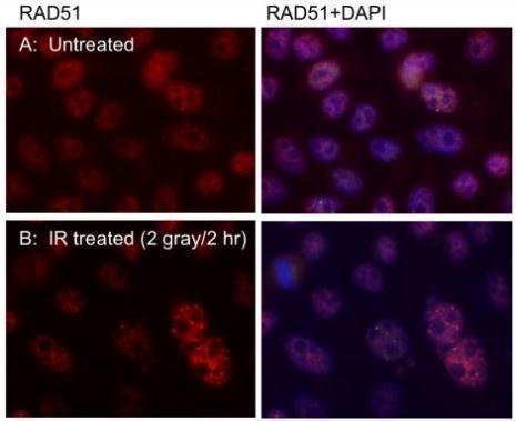 Immunofluorecent staning of RAD51 nuclear foci in U2OS cells using RAD51 14B4 antibody (56214).
Cells were pre-extracted with CSK buffer before fixation with 4% PFA.
RAD51 14B4 was used at 1:1000 dultion. DAPI was used to counterstain the nucleus.
Scale bar, 10 mciro-meter.
Immunofluorecent staning of RAD51 nuclear foci in U2OS cells using RAD51 14B4 antibody (56214).
Cells were pre-extracted with CSK buffer before fixation with 4% PFA.
RAD51 14B4 was used at 1:1000 dultion. DAPI was used to counterstain the nucleus.
Scale bar, 10 mciro-meter.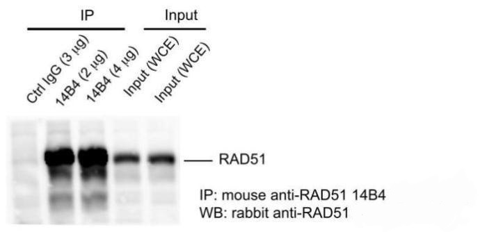 Mouse anti-RAD51 14B4 antibody (56214) was used in IP assay (immunoprecipitation) using HeLa cell extract prepared with lysis 180 buffer (40 mM Tris-HCl pH8.0, 180 mM NaCl, 1 mMEDTA, 0.5% NP-40).
Rabbit anti-RAD51 antibody was used for subsequent WB detection of immunoprecipated RAD51.
Whole cell extract was used as input.
Mouse anti-RAD51 14B4 antibody (56214) was used in IP assay (immunoprecipitation) using HeLa cell extract prepared with lysis 180 buffer (40 mM Tris-HCl pH8.0, 180 mM NaCl, 1 mMEDTA, 0.5% NP-40).
Rabbit anti-RAD51 antibody was used for subsequent WB detection of immunoprecipated RAD51.
Whole cell extract was used as input.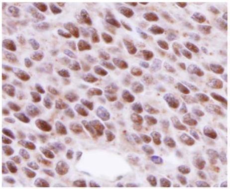 RAD51 antibody 14B4 (56214) detects RAD51 in nucleus on BT483 xenograft by immunohistochemical analysis.
Sample: Paraffin-embedded BT483 xenograft.
RAD51 antibody 14B4 (562148) dilution: 1:200.
Antigen Retrieval: Trilogy™ (EDTA based, pH 8.0) buffer, 15min
RAD51 antibody 14B4 (56214) detects RAD51 in nucleus on BT483 xenograft by immunohistochemical analysis.
Sample: Paraffin-embedded BT483 xenograft.
RAD51 antibody 14B4 (562148) dilution: 1:200.
Antigen Retrieval: Trilogy™ (EDTA based, pH 8.0) buffer, 15min![Various whole cell extracts (30 ug) were separated by 10% SDS-PAGE, and the membrane was blotted with Rad51 antibody [14B4] (56214) diluted at 1:500. The HRP-conjugated anti-mouset IgG antibody was used to detect the primary antibody, and the signal was developed with Trident ECL plus-Enhanced.](https://www.leinco.com/wp-content/uploads/2025/01/qed-bioscience-anti-rad51-antibody-56214-4.jpg) Various whole cell extracts (30 ug) were separated by 10% SDS-PAGE, and the membrane was blotted with Rad51 antibody [14B4] (56214) diluted at 1:500. The HRP-conjugated anti-mouset IgG antibody was used to detect the primary antibody, and the signal was developed with Trident ECL plus-Enhanced.
Various whole cell extracts (30 ug) were separated by 10% SDS-PAGE, and the membrane was blotted with Rad51 antibody [14B4] (56214) diluted at 1:500. The HRP-conjugated anti-mouset IgG antibody was used to detect the primary antibody, and the signal was developed with Trident ECL plus-Enhanced.![Various whole cell extracts (30 ug) were separated by 10% SDS-PAGE, and the membrane was blotted with Rad51 antibody [14B4] (56214) diluted at 1:500. The HRP-conjugated anti-mouset IgG antibody was used to detect the primary antibody, and the signal was developed with Trident ECL plus-Enhanced.](https://www.leinco.com/wp-content/uploads/2025/01/qed-bioscience-anti-rad51-antibody-56214-5.jpg) Various whole cell extracts (30 ug) were separated by 10% SDS-PAGE, and the membrane was blotted with Rad51 antibody [14B4] (56214) diluted at 1:500. The HRP-conjugated anti-mouset IgG antibody was used to detect the primary antibody, and the signal was developed with Trident ECL plus-Enhanced.
Various whole cell extracts (30 ug) were separated by 10% SDS-PAGE, and the membrane was blotted with Rad51 antibody [14B4] (56214) diluted at 1:500. The HRP-conjugated anti-mouset IgG antibody was used to detect the primary antibody, and the signal was developed with Trident ECL plus-Enhanced.![Whole cell extract (30 ug) was separated by 10% SDS-PAGE, and the membrane was blotted with Rad51 antibody [14B4] (56214) diluted at 1:500. The HRP-conjugated anti-mouse IgG antibody was used to detect the primary antibody.](https://www.leinco.com/wp-content/uploads/2025/01/qed-bioscience-anti-rad51-antibody-56214-6.jpg) Whole cell extract (30 ug) was separated by 10% SDS-PAGE, and the membrane was blotted with Rad51 antibody [14B4] (56214) diluted at 1:500. The HRP-conjugated anti-mouse IgG antibody was used to detect the primary antibody.
Whole cell extract (30 ug) was separated by 10% SDS-PAGE, and the membrane was blotted with Rad51 antibody [14B4] (56214) diluted at 1:500. The HRP-conjugated anti-mouse IgG antibody was used to detect the primary antibody.![Various whole cell extracts (30 ug) were separated by 10% SDS-PAGE, and the membrane was blotted with Rad51 antibody [14B4] (56214) diluted at 1:500. The HRP-conjugated anti-mouse IgG antibody was used to detect the primary antibody.](https://www.leinco.com/wp-content/uploads/2025/01/qed-bioscience-anti-rad51-antibody-56214-7.jpg) Various whole cell extracts (30 ug) were separated by 10% SDS-PAGE, and the membrane was blotted with Rad51 antibody [14B4] (56214) diluted at 1:500. The HRP-conjugated anti-mouse IgG antibody was used to detect the primary antibody.
Various whole cell extracts (30 ug) were separated by 10% SDS-PAGE, and the membrane was blotted with Rad51 antibody [14B4] (56214) diluted at 1:500. The HRP-conjugated anti-mouse IgG antibody was used to detect the primary antibody.![Rad51 antibody [14B4] detects Rad51 protein at nuclear foci by immunofluorescent analysis. Sample: Mock and treated HeLa cells were fixed in 4% paraformaldehyde at RT for 15 min. Green: Rad51 stained by Rad51 antibody [14B4] (56214) diluted at 1:500. Blue: Fluoroshield with DAPI.](https://www.leinco.com/wp-content/uploads/2025/01/qed-bioscience-anti-rad51-antibody-56214-8.jpg) Rad51 antibody [14B4] detects Rad51 protein at nuclear foci by immunofluorescent analysis.
Sample: Mock and treated HeLa cells were fixed in 4% paraformaldehyde at RT for 15 min.
Green: Rad51 stained by Rad51 antibody [14B4] (56214) diluted at 1:500.
Blue: Fluoroshield with DAPI.
Rad51 antibody [14B4] detects Rad51 protein at nuclear foci by immunofluorescent analysis.
Sample: Mock and treated HeLa cells were fixed in 4% paraformaldehyde at RT for 15 min.
Green: Rad51 stained by Rad51 antibody [14B4] (56214) diluted at 1:500.
Blue: Fluoroshield with DAPI. - -
- -
Antibody DetailsProduct DetailsReactive Species Human Host Species Mouse Immunogen GST fusion protein corresponding to full-length Rad51 (aa 1-338) of human Rad51 expressed in E. coli. Product Concentration Lot Specific Formulation PBS, pH 7.4 State of Matter Liquid Product Preparation Purified by affinity on GST Storage and Handling This antibody is stable for at least one (1) year at -70°C. Avoid multiple freeze- thaw cycles. Regulatory Status Research Use Only Country of Origin USA Shipping Next Day 2-8°C Applications and Recommended Usage? Quality Tested by Leinco Immunoblotting,
Immunoprecipitation: use at 1-5 ug/mL. Positive control: T24 bladder carcinoma cells and recombinant fusion protein. Each investigator should determine their own optimal working dilution for specific applications. See directions on lot specific datasheets, as information may periodically change. DescriptionSpecificity This antibody recognizes human Rad51, a 37 kD protein that interacts with BRCA1. This interaction is important for tumor suppression. BRCA2 and Rad51 have also been shown to interact via the BRC repeats located with exon 11 of BRCA2. When these repeats are deleted, the interaction is lost, and cells become hypersensitive to DNA damage caused by methyl methanesulfonate (MMS). Function Plays an important role in homologous strand exchange, a key step in DNA repair through homologous rPubMed:12205100, PubMed:12442171, PubMed:18417535, PubMed:20231364, PubMed:20348101, PubMed:20413593, PubMed:23509288, PubMed:23754376, PubMed:26253028, PubMed:26681308, PubMed:28575658}. NCBI Gene Bank ID UniProt.org Research Area Cancer Research References & Citations1. Shinohara, A et al. 1993. Nature Genetics 4: 239-243 2. Scully, R et al. 1997. Cell 88: 265-275 3. Chen, PL et al. 1998. PNAS (USA) 95: 5287-5292. Technical ProtocolsICC IF  IHC FFPE   Certificate of Analysis |
Formats Available
 Products are for research use only. Not for use in diagnostic or therapeutic procedures.